Le Infezioni in Medicina, n. 3, 355-360, 2026
CASE REPORTS
Aortic aneurysm and aorto-bi-iliac endoprosthesis infection caused by Helicobacter cinaedi diagnosed by next-generation sequencing: a case report
Arianna de Angelis1,2, Lucia Piazza1,2, Giulia la Martire2, Giulia Martelli2, Giorgio Dirani3, Vittorio Sambri1,3, Gianpiero Tebano1,2, Francesco Cristini1,2
1Department of Medical and Surgical Sciences (DIMEC), Alma Mater Studiorum, University of Bologna, Bologna, Italy;
2Infectious Diseases Unit, Forlì and Cesena Hospitals, AUSL Romagna, Forlì and Cesena, Italy;
3Unit of Microbiology, The Greater Romagna Area Hub Laboratory, AUSL Romagna, 47522 Cesena, Italy.
Article received 06 March 2026 and accepted 01 July 2026
Corresponding author
Gianpiero Tebano
E-mail: gianpiero.tebano@unibo.it
SUMMARY
Helicobacter cinaedi is an emerging Gram-negative pathogen, possibly involved in the pathogenesis of atherosclerosis and increasingly recognized as a cause of bacteraemia, cellulitis and vascular infections, particularly in immunocompromised and elderly patients. Infection of vascular grafts by H. cinaedi remains exceedingly rare. We report a case of H. cinaedi infection involving an aortic aneurysm and an aorto-bi-iliac endoprosthesis in an immunocompetent patient, highlighting the diagnostic challenges posed by culture-negative infections and the crucial role of Next-Generation Sequencing (NGS) in pathogen identification and therapeutic management.
Keywords: Helicobacter cinaedi, aortic aneurysm, aortic endoprosthesis, vascular infection, next-generation sequencing, 16S rRNA.
INTRODUCTION
Helicobacter cinaedi is a Gram-negative, spiral-shaped bacterium belonging to the enterohepatic Helicobacter group [1]. Among non–Helicobacter pylori Helicobacter (NHPH) species, H. cinaedi is increasingly recognized as a cause of human infection [2]. Its detection is difficult, as it rarely grows on standard culture media; when growth occurs, it is usually on enriched, non-selective media, under microaerophilic conditions. Therefore, molecular techniques such as 16S rRNA sequencing represent useful tools for its identification [3].
Helicobacter cinaedi is typically considered an opportunistic pathogen affecting immunosuppressed hosts; however, cases of infection in immunocompetent individuals have been described [4].
The organism most commonly causes bacteraemia, often accompanied by cellulitis (typically of the extremities) or nonspecific febrile illness characterized by fever, malaise, and fatigue. Recent reports have described an association between H. cinaedi and aortic aneurysm infections in immunocompetent patients [5]. Moreover, a possible role in promoting atherosclerosis through chronic infection has been proposed [6, 7]. We report a case of an aorto-bi-iliac endoprosthesis infection in which 16S rRNA NGS performed on periaortic tissue provided microbiological evidence of H. cinaedi infection.
CASE PRESENTATION
We report the case of a 73-year-old immunocompetent man. His past medical history included colonic diverticulosis and placement of an aorto-bi-iliac endoprosthesis in 2019 for an infrarenal aortic aneurysm.
The patient was referred for an infectious diseases (ID) consultation by the Rheumatology Unit for suspected aortitis and was then hospitalised in the ID ward. The condition that led to hospitalisation had begun approximately six months earlier (Figure 1). At that time, the patient underwent a follow-up contrast-enhanced computed tomography angiography (CTA), performed as part of routine surveillance of the endoprosthesis, which revealed mild mural enhancement and periaortic inflammatory fat stranding, suggestive of inflammation. To further investigate these findings, a fluorodeoxyglucose-positron emission tomography/computed tomography (FDG PET/CT) was performed, demonstrating focal metabolic uptake around the excluded aneurysmal sac and the adjacent endoprosthesis, suggestive of localized inflammation or infection. A vasculitic process was suspected, and the patient was treated with corticosteroids (oral prednisone, 25 mg daily for 1 week, then 12.5 mg daily, until the follow-up FDG PET/CT).
A follow-up FDG PET/CT was performed 6 weeks later and revealed increased metabolic activity (Maximum Standardized Uptake Value - SUVmax - 18.25 vs. 11.8), despite treatment with corticosteroids, raising a strong suspicion of graft infection (Figure 2) and leading to the hospitalisation in our ID ward.
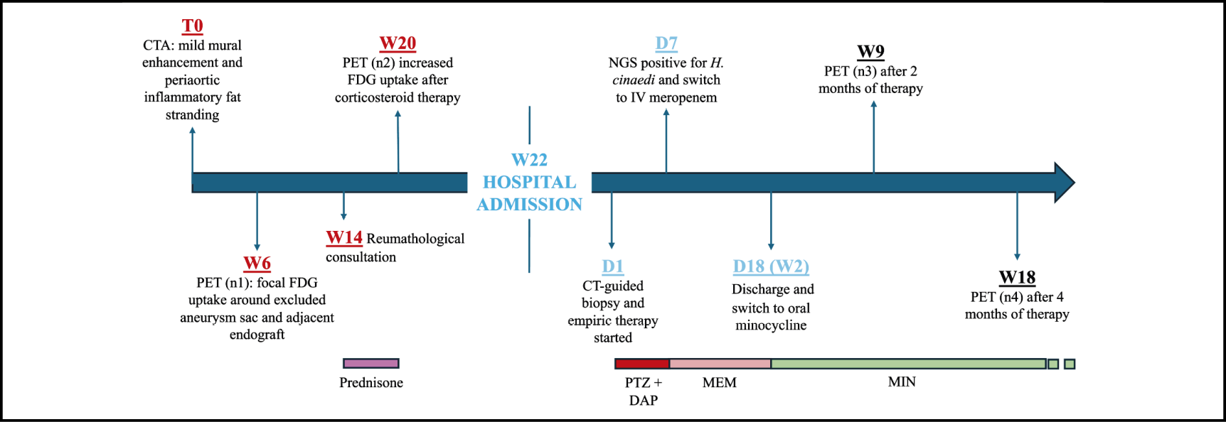
Figure 1 - Clinical timeline of diagnosis, microbiological workup and treatment of Helicobacter cinaedi endograft infection. Note: CT: computed tomography; CTA: computed tomography angiography; D: days after admission; DAP: daptomycin; FDG: Fluorodeoxyglucose; M: month; MEM: meropenem; NGS: next-generation sequencing; PET: Positron Emission Tomography; PTZ: piperacillin/tazobactam; W: weeks before/after admission.
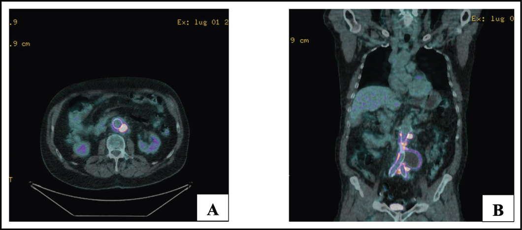
Figure 2 - Baseline 18F-FDG PET/CT performed at the time of diagnosis. Axial (A) and coronal (B) fused PET/CT images demonstrate intense FDG uptake along the abdominal aortic endograft and surrounding periaortic tissue, with a SUVmax of 13.6.
On admission, the patient was afebrile and reported mild, diffuse arthromyalgias as the only relevant symptom. He had had elevated inflammatory markers for at least six months. The patient was evaluated by vascular surgeons to discuss surgical removal of the endoprosthesis, to achieve complete source control of the infection. The surgical option was judged unfeasible by surgeons, due to an excessive intra- and postoperative risk.
Consequently, a CT-guided biopsy of the periaortic adipose tissue surrounding the aneurysmal sac was performed, targeting the area with increased metabolic activity on FDG PET/CT. Empiric antibiotic therapy with piperacillin/tazobactam 4.5 g four times daily and daptomycin 850 mg once daily was then initiated while awaiting microbiological results.
Blood and tissue cultures showed no growth. Therefore, 16S rRNA next-generation sequencing (NGS) of the biopsy specimen was performed. The biopsy specimen was subjected to pretreatment by mechanical lysis using 0.5 mm PowerBead Tubes Glass (Qiagen) and TissueLyser II (Qiagen). Nucleic acid extraction was performed using the Maxwell® CSC Pathogen Total Nucleic Acid Kit (Promega) on the Maxwell 48 CSC automated platform (Promega). The resulting eluate was subsequently processed for library preparation using the Microbiota Solution A™ kit (AD4Seq, Arrow Diagnostics), which enables the amplification and sequencing of the V1–V2–V3 hypervariable regions of the 16S rRNA gene. Sequencing was performed on the Illumina MiSeq platform using the MiSeq Reagent Nano Kit v2 (500 cycles).
Bioinformatic analysis was carried out using the DRAGEN 16S Plus application (Illumina BaseSpace), which employs the NCBI RefSeq RDP database version 1 for processing of raw FASTQ files and taxonomic assignment of reads. The total number of reads assigned to the domain Bacteria was 1,523, of which 1,129 were assigned to Helicobacter cinaedi, corresponding to a relative abundance of 74%. This value is substantially above the method’s positivity threshold, set at 100 reads. In the negative control processed in parallel, no reads attributable to H. cinaedi were detected. No confirmatory PCR or Sanger sequencing targeting H. cinaedi was performed. A selective culture for Helicobacter spp. was also performed on PYL agar (bioMérieux) under microaerophilic conditions at 37°C for 10 days, with negative results.
After 7 days of empiric therapy, once the NGS results became available, targeted treatment with intravenous meropenem was initiated and administered for an additional 10 days (1 g three times daily), followed by oral minocycline (100 mg twice daily) after hospital discharge.
In the following months, the patient remained asymptomatic. He reported good adherence to antibiotic treatment with minocycline, without significant side effects.
FDG PET/CT was repeated 2 months and 4 months after initiation of antibiotic therapy and showed a progressive reduction in metabolic activity within the infected areas (SUVmax 13.6 and SUVmax 10.9, respectively) (Figure 3). The patient also underwent a follow-up CTA, confirming the absence of endograft leaks.
At the time of writing, the patient is still undergoing treatment with minocycline twice daily, as chronic suppressive therapy. New FDG PET/CT and CTA have been scheduled for the 1-year follow-up. The duration of antibiotic therapy will be adjusted according to clinical and radiological findings and treatment tolerability.
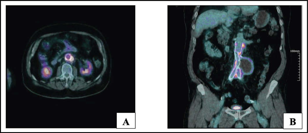
Figure 3 - Follow-up 18F-FDG PET/CT performed 4 months after initiation of antimicrobial therapy. Axial (A) and coronal (B) fused PET/CT images show persistent FDG uptake along the abdominal aortic endograft and surrounding periaortic tissue, with a lower SUVmaxx compared with baseline (10.6 vs 13.6).
DISCUSSION
Infected aortic aneurysms due to H. cinaedi are rare but increasingly recognized in both immunocompetent and immunocompromised patients [7]. Early reports described H. cinaedi almost exclusively in immunocompromised patients, such as people living with HIV or agammaglobulinemia, leading to the assumption that the organism was associated with specific host conditions [8, 9]. However, an increasing number of H. cinaedi infections in immunocompetent patients have been reported more recently, showing that the group of patients at risk may be larger than originally thought [5, 10, 11]. Our patient had no immunosuppressive conditions, as he had negative HIV serology, no malignancy, and no ongoing immunosuppressive therapy. Although the patient received a short course of corticosteroid therapy, it was administered at low doses, below those conventionally considered defining a state of immunosuppression. Moreover, the clinical condition was already present at that time.
The infection of the aortic aneurysm and aorto-bi-iliac endoprosthesis was probably preceded by H. cinaedi bacteraemia, since no contiguous infectious foci were identified. Assuming this hypothesis, the source of bacteraemia remains uncertain. Given the patient’s known diverticulosis, bacterial translocation from the intestinal tract seems to be the most plausible mechanism, as already suggested by other authors [12, 13].
As in previously reported H. cinaedi aortic infections, microbiological diagnosis was challenging because of the fastidious nature of the organism, requiring prolonged incubation in automated blood culture systems [14]. Even with prolonged incubation, detection rates remain low [7]. Moreover, in 23.3%–25% of cases of aortic aneurysm infections, the causative pathogen remains unidentified [15].
In our case, both routine blood cultures and extended cultures on appropriate enriched media from the tissue specimen were negative. Next-generation sequencing proved essential, suggesting the value of culture-independent molecular diagnostics for identifying fastidious organisms and guiding pathogen-specific therapy [16]. The added value of molecular diagnostics in vascular graft infections has been demonstrated in previous studies, where sequencing of intraoperative tissue and explanted graft specimens, combined with conventional cultures, improved pathogen detection compared with culture alone. This approach may be particularly useful in patients exposed to antimicrobial therapy before sampling, in whom conventional cultures are often negative [17]. Similarly, in our patient, NGS performed on periaortic tissue enabled identification of H. cinaedi despite negative conventional microbiological investigations.
Notably, pathogen identification is a major criterion for aortic graft infection, according to the Management of Aortic Graft Infection Collaboration (MAGIC) criteria [18]. In addition, one major radiological criterion and one minor radiological criterion were fulfilled (i.e. CT and PET findings, respectively), together with one minor laboratory criterion, namely elevated serum C-reactive protein (CRP) levels. These findings, therefore, allowed a definitive diagnosis, which until then was only suspected from imaging.
The identification of H. cinaedi enabled optimization of antibiotic therapy, including de-escalation to a narrower-spectrum agent (minocycline). The optimal treatment for infection of aortic aneurysm caused by H. cinaedi is unknown, in terms of intravenous (IV) vs oral therapy and with respect to treatment duration. Some authors have suggested an approach based on IV antibiotics (at least 6 weeks), while others have proposed a prolonged course of oral antimicrobials, ranging from 6 months to lifelong therapy. Considering the limited and conflicting evidence currently available, we decided to initiate a 10-day course of intravenous meropenem as induction therapy, with the aim of ensuring high-dose antimicrobial exposure through a drug capable of achieving high intravascular concentrations. This was subsequently followed by continuation therapy with a tetracycline (minocycline), given that this class of agents is characterized by favourable tissue penetration and is widely employed in chronic suppressive regimens in the setting of osteoarticular infections. Since NGS does not provide information regarding antimicrobial susceptibility, in both instances the therapeutic choice was guided by data available in the literature on susceptibility rates to these agents, which appear to be favourable. H. cinaedi strains generally exhibit low minimum inhibitory concentration (MIC) values for carbapenems, aminoglycosides, and tetracycline (MIC90≤1 mg/L for imipenem, gentamicin, and tetracycline). In contrast, penicillins and cephalosporins display moderate MIC values (MIC90=16 mg/L for ampicillin and carbenicillin, and MIC90=8 mg/L for amoxicillin, cefepime, and ceftriaxone) [14].
Moreover, intravascular device removal and replacement remain imperative when feasible [7]. In this case, the removal of the endoprosthesis was judged unfeasible by the surgeons, because of excessive surgical risk. Therefore, a prolonged course of treatment (suppressive therapy) has been planned, considering the absence of source control as a major risk for relapse if antibiotic treatment is discontinued.
Presently, this therapeutic approach has been accompanied by clinical improvement and a reduction in metabolic activity on FDG PET/CT imaging. However, given the persistence of prosthetic material and the limited duration of follow-up, the long-term effectiveness of this approach remains uncertain.
Our patient is currently being monitored through imaging and laboratory assessments to evaluate the regression of the infectious involvement of the aortic aneurysm and aortic endoprosthesis. Based on the tolerability of the antibiotic and the evolution of FDG PET/CT imaging, we will decide whether to continue treatment after the 1-year follow-up.
In conclusion, H. cinaedi should be considered in the differential diagnosis of culture-negative vascular graft infections, even in immunocompetent patients. Although vascular or endograft infections caused by H. cinaedi are exceedingly rare, the relevance of the present report may extend beyond the rarity of the pathogen itself. The case illustrates how NGS may provide microbiological evidence contributing to a confirmed diagnosis of endograft infection.
Acknowledgments
A preliminary report of this case was presented at the National Congress of the Italian Society of Infectious Diseases and Tropical Medicine (SIMIT) 2025, poster number 177.
Ethics
The patient provided his informed consent to the description of this case report.
Conflict of Interest
The authors have no relevant financial or non-financial interests to disclose.
Funding
The authors declare that no funds, grants, or other support were received during the preparation of this manuscript.
REFERENCES
[1] Gotoh Y, Atsuta Y, Taniguchi T, et al. Helicobacter cinaedi is a human-adapted lineage in the Helicobacter cinaedi/canicola/‘magdeburgensis’ complex. Microb Genom. 2022; 8(5): 000830.
[2] Fox-Lewis A, Basu I, Vesty A, Henderson G, Chhibber AV, Thomas M. Helicobacter cinaedi bacteremia in a returning traveler. IDCases. 2020; 21: e00910
[3] Matsumoto T, Goto M, Murakami H, et al. Multicenter study to evaluate bloodstream infection by Helicobacter cinaedi in Japan. J Clin Microbiol. 2007; 45(9): 2853-2857
[4] Bateman AC, Butler-Wu SM. The Brief Case: bacteremia caused by Helicobacter cinaedi. J Clin Microbiol. 2017; 55(1): 5-9.
[5] Gorak Savard R, Savoie-White FH, Gegiia I, et al. Helicobacter cinaedi as a cause of primary aortic infections and the challenges of diagnosis and optimal treatment. J Vasc Surg Cases Innov Tech. 2025; 11(3): 101744.
[6] Matsuoka A, Sasaki Y, Kubodera A, et al. Acquired hemophilia A presenting with infectious aortic aneurysms due to an underlying Helicobacter cinaedi infection. Intern Med. 2021; 60(24): 3947-3952.
[7] Matsuo T, Mori N, Mizuno A, et al. Infected aortic aneurysm caused by Helicobacter cinaedi: case series and systematic review of the literature. BMC Infect Dis. 2020; 20(1): 854.
[8] Tee W, Street AC, Spelman D, Munckhof W, Mijch A. Helicobacter cinaedi bacteraemia: varied clinical manifestations in three homosexual males. Scand J Infect Dis. 1996; 28(2): 199-203.
[9] Simons E, Spacek LA, Lederman HM, Winkelstein JA. Helicobacter cinaedi bacteremia presenting as macules in an afebrile patient with X-linked agammaglobulinemia. Infection. 2004; 32(6): 367-368.
[10] Murata S, Suzuki H, Sakamoto S, et al. Helicobacter cinaedi-associated vertebral osteomyelitis in an immunocompetent patient. Intern Med. 2015; 54(24): 3221-3224.
[11] Shimizu Y, Gomi H, Ishioka H, Isono M. Refractory to treat Helicobacter cinaedi bacteremia with bilateral lower extremities cellulitis in an immunocompetent patient. IDCases. 2016; 5: 9-11.
[12] Araoka H, Baba M, Okada C, et al. First evidence of bacterial translocation from the intestinal tract as a route of Helicobacter cinaedi bacteremia. Helicobacter. 2018; 23(1): e12458.
[13] Araoka H, Baba M, Kimura M, et al. Clinical characteristics of bacteremia caused by Helicobacter cinaedi and time required for blood cultures to become positive. J Clin Microbiol. 2014; 52(5): 1519-1522.
[14] Kawamura Y, Tomida J, Morita Y, et al. Clinical and bacteriological characteristics of Helicobacter cinaedi infection. J Infect Chemother. 2014; 20(9): 517-526.
[15] Sörelius K, Budtz-Lilly J, Mani K, Wanhainen A. Systematic review of the management of mycotic aortic aneurysms. Eur J Vasc Endovasc Surg. 2019; 58(3): 426-435.
[16] Nafea AM, Wang Y, Wang D, et al. Application of next-generation sequencing to identify different pathogens. Front Microbiol. 2024; 14: 1329330.
[17] Ajdler-Schaeffler E, Scherrer AU, Keller PM, et al. Increased pathogen identification in vascular graft infections by the combined use of tissue cultures and 16S rRNA gene polymerase chain reaction. Front Med (Lausanne). 2018; 5: 169.
[18] Anagnostopoulos A, Mayer F, Ledergerber B, et al. Editor’s Choice - Validation of the Management of Aortic Graft Infection Collaboration (MAGIC) criteria for the diagnosis of vascular graft/endograft infection: results from the prospective vascular graft cohort study. Eur J Vasc Endovasc Surg. 2021; 62(2): 251-257.