Le Infezioni in Medicina, n. 3, 324-333, 2026
ORIGINAL ARTICLES
Environmental isolation and in vitro characterisation of bacteriophages active against multidrug-resistant Acinetobacter baumannii
Souradeep Chowdhury¹, Nisha Rathor², Manish Soneja³, Bimal Kumar Das², Ranveer Singh Jadon³, Naveet Wig³, Rama Chaudhry²
¹Department of Medicine and Microbiology, All India Institute of Medical Sciences (AIIMS), New Delhi, India;
²Department of Microbiology, All India Institute of Medical Sciences (AIIMS), New Delhi, India;
³Department of Medicine, All India Institute of Medical Sciences (AIIMS), New Delhi, India.
Article received 20 March 2026 and accepted 08 July 2026
Corresponding author
Rama Chaudhry
E-mail: drramach@gmail.com
SUMMARY
Background: The increasing prevalence of antimicrobial resistance (AMR) has created an urgent need for alternative therapeutic strategies. Acinetobacter baumannii, a major cause of hospital-acquired infections, has been classified by the World Health Organization as a critical priority pathogen due to its high levels of multidrug resistance. Bacteriophage therapy has re-emerged as a potential approach to combat infections caused by multidrug-resistant organisms.
Methods: Fifty multidrug-resistant (MDR) A. baumannii clinical isolates were collected. Bacteriophages were isolated from hospital sewage using standard enrichment protocols and screened for lytic activity against these isolates using spot and plaque assays. Morphological characterization of purified phages was performed using transmission electron microscopy.
Results: Three bacteriophages – vB_AbaS_SRNAIIMS002, vB_AbaS_SRNAIIMS008, and vB_AbaP_SRNAIIMS010 – were isolated and demonstrated lytic activity against MDR A. baumannii. Plaque assays revealed clear central zones with surrounding halos, indicating active bacterial lysis. Host range testing showed that two phages exhibited activity against multiple clinical isolates, whereas one displayed a narrow host range. Transmission electron microscopy revealed that two phages belonged to the family Siphoviridae, whereas one was morphologically consistent with the family Podoviridae.
Conclusions: This study reports the isolation and characterization of three bacteriophages active against MDR A. baumannii. These findings highlight the feasibility of isolating lytic phages from environmental sources and support further investigation of bacteriophage therapy as a complementary strategy to address infections caused by multidrug-resistant pathogens.
Keywords: Bacteriophage therapy, Acinetobacter baumannii, multidrug resistance, phage isolation, antimicrobial resistance
INTRODUCTION
The widespread use of antimicrobials has played a pivotal role in enabling many of the advances in modern medicine, including complex surgical procedures, intensive care and oncologic interventions. However, the increasing prevalence of infections caused by multidrug-resistant organisms (MDROs) has emerged as a significant barrier to effective clinical care. Among these, Acinetobacter baumannii has gained particular prominence due to its extensive drug resistance and association with high morbidity and mortality in hospital settings [1, 2]. Indeed, in the 2017 WHO list of organisms against which new antibiotic development is urgently required, the name of Acinetobacter baumannii was listed among the critical-priority pathogens [3].
The limited therapeutic options for this pathogen have prompted renewed interest in alternative antimicrobial strategies.
One such strategy is bacteriophage therapy, which involves the use of viruses that specifically infect and lyse bacteria. While the therapeutic potential of bacteriophages was recognized as early as 1919, interest waned following the advent of broad-spectrum antibiotics [4]. With rising resistance trends, bacteriophage therapy is re-emerging as a promising area of research [5].
This study aimed to isolate and characterise novel bacteriophages targeting A. baumannii, with the objective of evaluating their host range, morphological characteristics, and in vitro lytic activity to determine their therapeutic potential. An overview of the overall study workflow is shown in Figure 1.
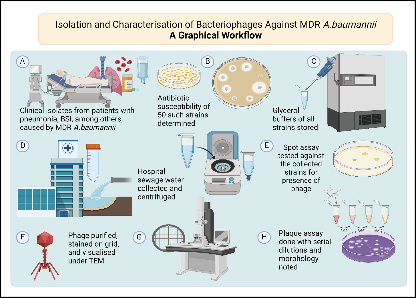
Figure 1 - Graphical overview of bacteriophage isolation, characterisation, and lytic activity assessment against multidrug-resistant Acinetobacter baumannii.
MATERIALS AND METHODS
Bacterial isolate identification and susceptibility testing
The work was conducted between January 2020 and June 2022 following ethical clearance from the Institute Ethical Committee (Ref. No. IECPG-636 / 28.11.2019).
Fifty clinical isolates of Acinetobacter baumannii were obtained from the Bacteriology Lab, Department of Microbiology. Species identification was performed using the MALDI-TOF system and subsequent susceptibility testing was done by disc diffusion method in accordance with the Clinical and Laboratory Standards Institute (CLSI) guidelines [6]. Isolates were classified as MDR if they were resistant to at least one agent in three or more antimicrobial classes. Antibiotic classes tested included beta-lactam/beta-lactamase inhibitor combinations (piperacillin-tazobactam), carbapenems (imipenem, meropenem), fluoroquinolones (ciprofloxacin), cephalosporins (ceftazidime), aminoglycosides (amikacin), and polymyxins (colistin). Isolates with intermediate results were categorised as non-susceptible, in accordance with CLSI breakpoints [7].
Isolation, purification and host range determination of bacteriophages
Hospital sewage water was collected, maintaining standard precautions, and transported to the laboratory. The isolation of bacteriophages (henceforth interchangeably referred to as phages) was done according to the protocol described by Ellis et al., with slight modifications [8]. Briefly, the sewage water was treated with 1% chloroform (v/v) and centrifuged at 10,000 rpm for 10 minutes. The resultant supernatant was mixed 1:1 by volume with the log-phase bacterial cultures described above, and incubated overnight at 37°C. This was similarly treated with chloroform and after centrifugation, the supernatant was used for spot and plaque assays for phage detection and host range assessment. The details of the spot assay are given in Figure 2.
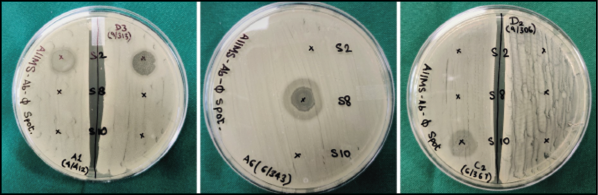
Figure 2 - Spot assay of the isolated bacteriophages against selected strains.
To observe the lytic activity of bacteriophages, bacterial suspensions adjusted to an OD600 of 2.0 were plated on Mueller-Hinton Agar (MHA) (HiMedia Laboratories Pvt. Ltd) and incubated at 37°C for 2 hours. A 10 µL aliquot of the phage preparation was spotted onto the bacterial lawn and incubated overnight to observe zones of lysis.
For the plaque assay, serial dilutions (10-fold) were made from bacteriophage preparation and 100 μL from each dilution was added to the exponentially grown host bacterial culture (200 μL), and incubated for 15 min at 37°C. To the mixture, 5 mL of molten soft agar (0.75% agar) was added and overlaid onto prepared MHA or LB agar plate (with 1.5% agar). The plates were incubated at 37°C overnight to observe the plaques. Both assays were performed according to the method described by Ellis et al., with slight modifications [8].
Transmission electron microscopy
Purified bacteriophage particles (500 μL) (107-108PFU/mL) were filtered using a 0.22 μm membrane filter, centrifuged at 25,000 x g for 75 min, and the pellet was washed with 0.1 M ammonium acetate (pH 7.0). The resuspended pellet wasloaded onto a copper grid for 1 min followed by negative staining with 1% uranyl acetate (pH 7.0) and drying. The method that was followed was that of Newase et al., with slight modifications [9]. The stained grid was visualized under Transmission Electron Microscopy (Thermo Scientific Talos L120C TEM) at an accelerating voltage of 120 kV. Virus particle dimensions were measured using the ImageJ computer program (version 1.53e), with the software scale calibrated using the scale bar obtained from the electron micrographs.
Bacteriophage lytic activity
Bacteriophage kinetics was performed to study the in vitro lysis of bacteria through a change in absorbance of optical density. The method followed was modified from that of Chen et al. [10] Briefly, 180 µL of bacterial culture in the log phase (105 CFU/mL) was aliquoted into wells of a 96-well microtiter plate. 20 µL of bacteriophage suspensions was added to wells at different multiplicities of infection (MOIs) (100, 10, 1, 0.1, 0.01, 0.001). MHA broth without Acinetobacter baumannii was used as a negative control, and A. baumannii bacteria without phage was a positive control for the experiment. The microtiter plate was incubated at 37°C inside an incubator, and change in absorbance at OD600 was recorded at 10 min intervals for 390 min. Each experiment was performed in triplicate.
RESULTS
Bacterial isolates
A total of 50 clinical isolates were obtained from clinical specimens. As shown in Figure 3, the majority were from respiratory sources: endotracheal aspirates (n=32), sputum (n=6), and bronchoalveolar lavage (BAL) fluid (n=4). Other isolates were from sterile body fluids (n=2), cerebrospinal fluid (n=1), blood (n=1), and wound-related samples, including tissues, swabs, and drain fluid.
All isolates met the criteria for multidrug resistance, showing non-susceptibility to agents in three or more antimicrobial classes (Figure 4). Notably, 40 of the 50 isolates (80%) were resistant to carbapenems. High resistance rates were also observed for piperacillin/tazobactam (90%), ciprofloxacin (96%), ceftazidime (96%), and amikacin (94%). All isolates tested for colistin were susceptible by broth microdilution.
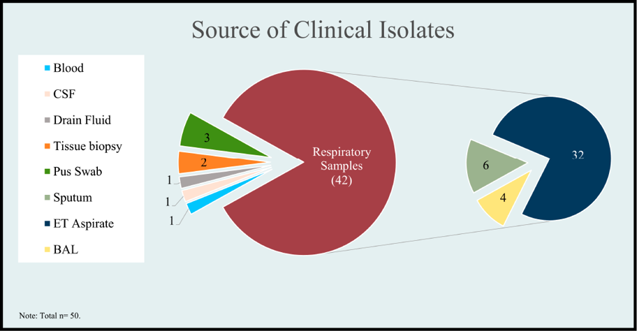
Figure 3 - Pie chart showing the various sources of the clinical isolates.
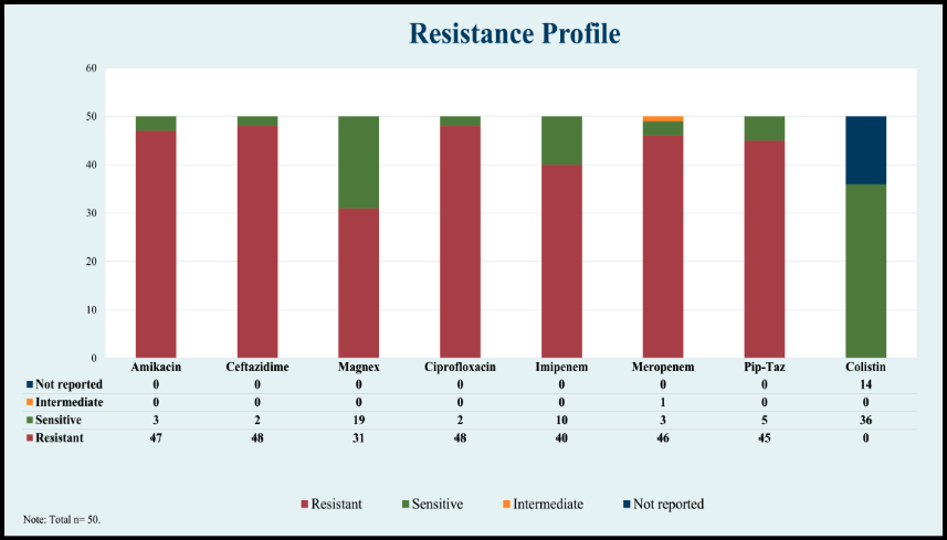
Figure 4 - The resistance profile of the Acinetobacter baumannii isolates.
Bacteriophage morphology and host range
A total of three bacteriophages were isolated from the sewage water, designated as vB_AbaS_SRNAIIMS002, vB_AbaS_SRNAIIMS008 and vB_AbaP_SRNAIIMS010. The bacteriophages were named in accordance with the proposed nomenclature for bacteriophages by Kropinski et al. For brevity, the phages are referred to as SRNAIIMS002, SRNAIIMS008 and SRNAIIMS010 respectively [11].
The plaque assay, done for all 3 phages with increasing dilutions, showed similar morphology and size of plaques with surrounding halos, confirming the presence of pure phage. SRNAIIMS002 showed a clear central zone measuring 3-4 mm in diameter. with a 1-2mm radial halo around it. For phage SRNAIIMS008, the central clearing measured 2-3mm in diameter, with a larger halo measuring 4-5mm radially. SRNAIIMS010 had a 1-2mm clear zone and a surrounding 2-3mm halo (Figure 5).
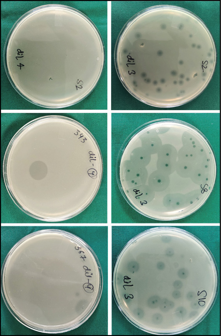
Figure 5 - Photograph showing plaque assay of the three isolated bacteriophages.
The spot assay was performed to evaluate the host range of the three isolated phages. Phage SRNAIIMS008 showed lytic activity only against its parent strain (1/50 isolates; 2%). Phage SRNAIIMS002 lysed 5/50 isolates (10%), and phage SRNAIIMS010 lysed 11/50 isolates (22%). Combined, the three phages demonstrated activity against 17/50 isolates (34%). Lysis was confirmed by both spot and plaque assays. The details of the host range are given in Table 1.
Table 1 - Isolated bacteriophages and their host range.

The morphologies of the isolated bacteriophages were revealed by TEM. For Phage SRNAIIMS002, the head measured 76.15 nm across while the tail was 245.82 nm long (Figure 6). For Phage SRNAIIMS008, the head measured 43.5 nm across while the tail was 97.38 nm long (Figure 7). The morphology of both phages was consistent with the family Siphoviridae of order Caudovirales, based on head-tail architecture; however, definitive taxonomic classification requires genomic characterisation, which was not performed in this study. Phage SRNAIIMS010 had a prominent icosahedral head measuring 47.46 nm across and a short stubby tail, morphologically consistent with the family Podoviridae of Order Caudovirales (Figure 8).
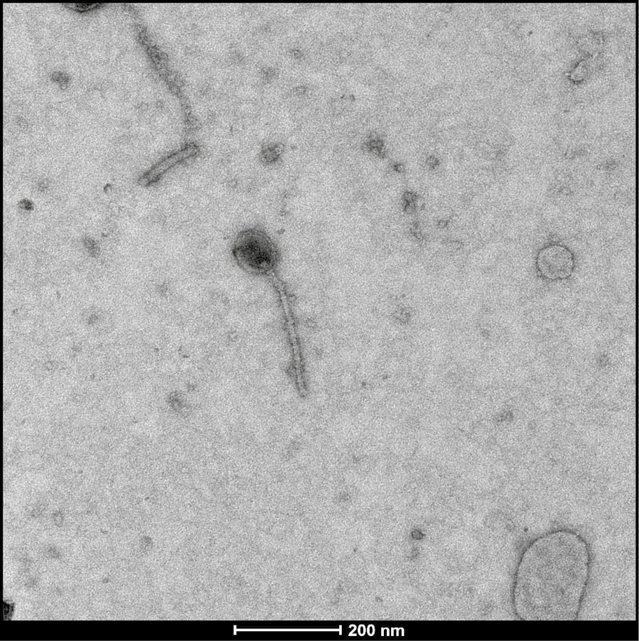
Figure 6 - TEM image of Phage vB_AbaS_SRNAIIMS002, a bacteriophage of the family Siphoviridae, with a head measuring 76.15 nm across and a 245.82 nm long tail.
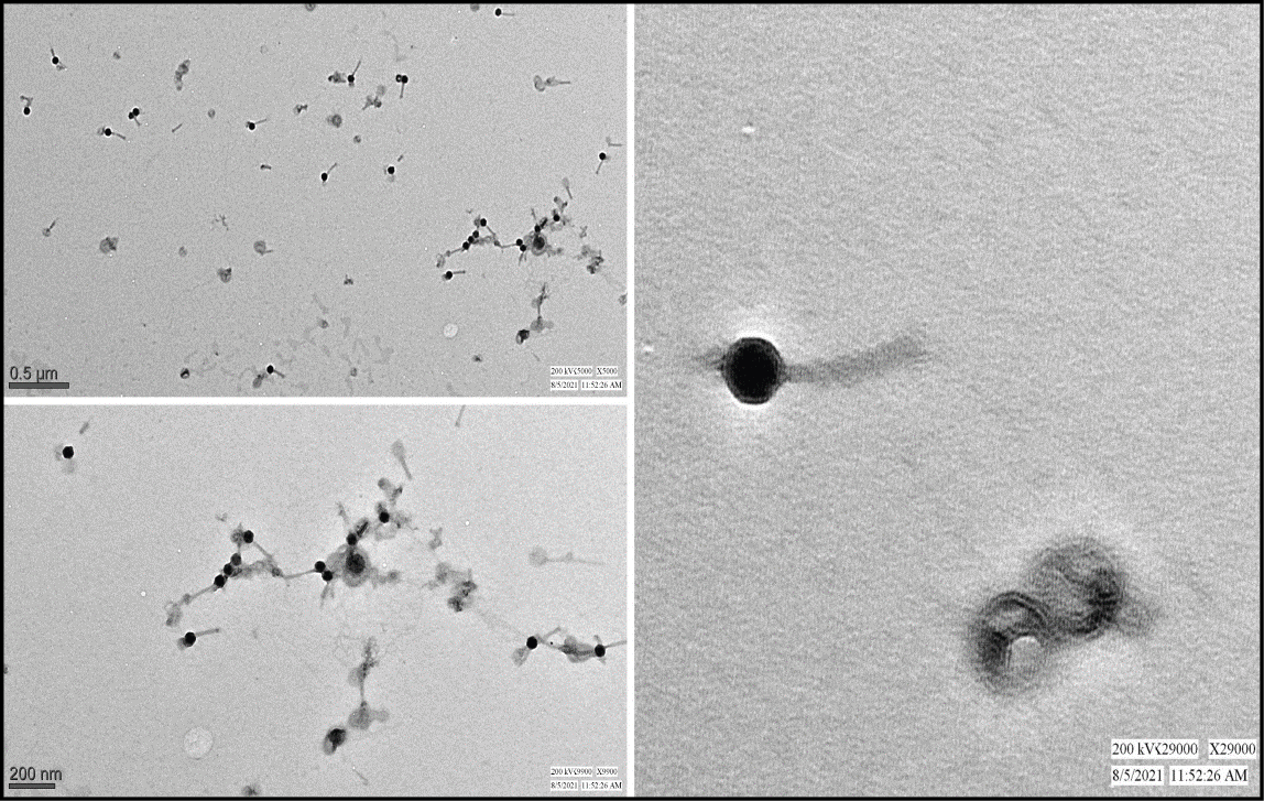
Figure 7 - TEM image of Phage vB_AbaS_SRNAIIMS008, a bacteriophage of the family Siphoviridae, with a head measuring 43.5 nm across and a 97.38 nm long tail.
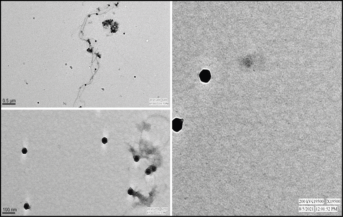
Figure 8 - TEM image of Phage vB_AbaP_SRNAIIMS010, a bacteriophage of the family Podoviridae, with a head measuring 47.46 nm across, and a short and stubby tail.
Phage lytic activity in vitro
Phage action on bacterial growth was observed through a change in OD600 over 8 h of incubation. In control wells (MOI: 0), uninhibited bacterial growth followed a typical sigmoidal curve. In contrast, wells treated with phages at various MOIs (100 to 0.001) demonstrated varying degrees of bacterial growth inhibition, confirming phage-mediated lysis (Figure 9a-c). Higher MOIs (100 and 10) consistently produced the most pronounced and sustained suppression of bacterial growth across all three phages. At lower MOIs (0.01 and 0.001), partial or delayed bacterial regrowth was observed after an initial period of lysis, indicating that bacterial populations can partially recover at suboptimal phage-to-bacterium ratios. These findings confirm that lytic efficacy is MOI-dependent in vitro.
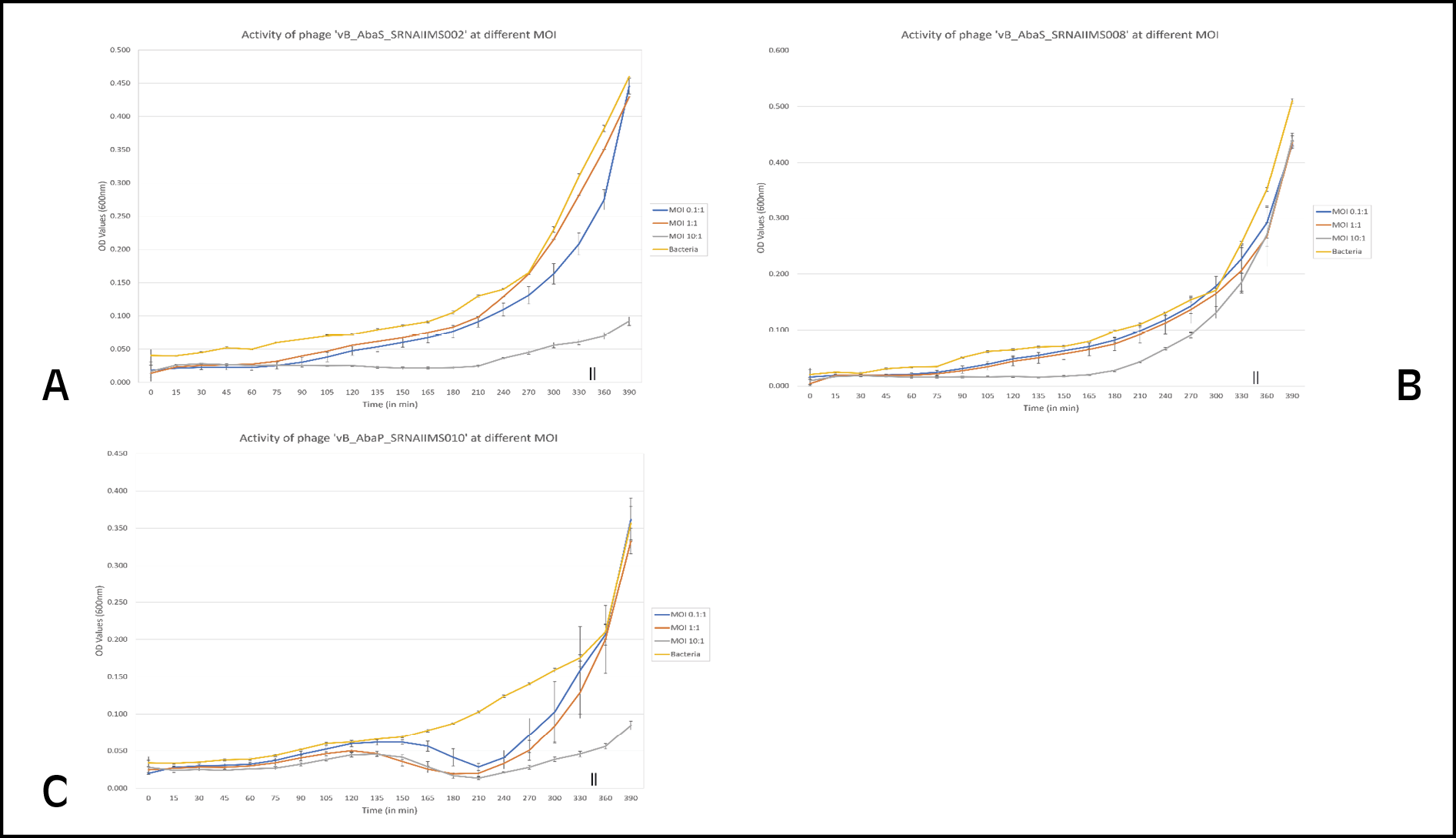
Figure 9 - Phage activity experiments for the three bacteriophages at different MOI. a) Phage vB_AbaS_SRNAIIMS002. b) Phage vB_AbaS_SRNAIIMS008. c) Phage vB_AbaP_SRNAIIMS010.
DISCUSSION
Acinetobacter baumannii represents an important cause of hospital-acquired infections, especially among hospitalized patients in the ICU [12]. The most common A. baumannii infections in critically ill patients include VAP, bloodstream infections (BSI), complicated urinary tract infections (cUTI), and wound infections [13]. An important characteristic of A. baumannii is its remarkable ability to persist in the environment and the rapid spread across wards [12]. Furthermore, A. baumannii is not only intrinsically resistant to multiple antimicrobials but also prone to acquiring new resistance determinants through a variety of different mechanisms. [12, 14] In the last 10 years, Acinetobacter baumannii has emerged as one of the major pathogens implicated in outbreaks of hospital-acquired infections, especially in patients from intensive care units [15-17]. Historically, carbapenems have been the most potent and reliable β-lactam antibiotics for the treatment of serious infections caused by A. baumannii [12]. However, the prevalence of carbapenem-resistant A. baumannii (CRAB) is seriously compromising the use of carbapenems in the control of such infections [18]. CRAB is usually resistant to almost all available antimicrobials. Although colistin is effective against most CRAB, it is a last-resort treatment due to high toxicity. Consequently, treatment options for these infections are limited and considerable mortality is associated with CRAB (about 50%) [19]. According to CHINET surveillance from China, the average resistance rate of A. baumannii to carbapenems is more than 70% [20]. In India too, the worrying rates of resistance in Acinetobacter baumannii were highlighted by data from the Indian Council of Medical Research (ICMR) [21]. These findings highlight the urgent medical need to promote alternatives to fight against CRAB infections.
Novel therapies including bacteriophage therapy against drug-resistant A. baumannii have been reported and reviewed [22-24]. There have been multiple attempts to use bacteriophages both in animal models and as instruments of infection control [25-27].
Successful phage control of various strains of Acinetobacter was demonstrated not only in vitro but also in vivo [28]. Two newly isolated phages infecting A. baumannii were characterized and suggested as potential candidates for phage cocktail [29, 30]. Other two newly isolated phages were characterized at genomic DNA level and suggested as potential candidates for phage cocktail against CRAB [31]. Phage Bϕ-C62 was used to successfully control CRAB infection the intranasal route in mice model [32]. In addition to demonstrating that phage treatment can be efficacious when a panel of phages is isolated from the environment against the particular strain causing the infection, an animal study has also demonstrated that the surviving A. baumannii bacteria had decreased virulence [33]. A combined lysis spectrum of four lytic phages against clinically isolated CRAB was reported to be 87.5% and phages were proven effective as therapeutic agents for lung infection without deleterious side effects in mice model [34]. Phages from multi-institute libraries were used to make a personalized cocktail and proven effective for treating a diabetic human patient with necrotizing pancreatitis complicated by an MDR A. baumannii infection [35]. A bacteriophage-containing aerosol was proven effective for cleaning and decreased the rates of infection caused by CRAB in intensive care units [36].
To our knowledge, this is among the few studies from India to report the isolation and in vitro characterisation of lytic bacteriophages from hospital sewage specifically targeting MDR A. baumannii clinical isolates. While previous studies have characterised A. baumannii phages from international sources, data from Indian clinical settings, where carbapenem resistance rates frequently exceed 80%, remain limited. This study adds to the growing body of evidence supporting environmental phage isolation as a strategy for identifying candidates for future therapeutic development. In this work, a total of 3 phages were specifically isolated for A. baumannii from hospital sewage, two of which exhibited strong efficiency at lysing different A. baumannii clinical isolates, while one had a narrower host range. This suggests that a variety of lytic A. baumannii phages can be readily isolated from the environment. These preliminary findings suggest potential future application in controlling clinical and hospital-acquired infections, although further validation, including genomic characterisation, in vivo studies, and safety assessment, will be required before any clinical translation can be considered. Two of the phages isolated were morphologically consistent with the family Siphoviridae, while one was consistent with the family Podoviridae.
Among the merits of this study, this was a treatment research study, and pertains directly to the work in the field of non-antimicrobial methods of treatment that is being carried out currently. The phages isolated in the course of our study can be sent to phage banks for storage. In the context of Indian studies, this type of research is being done in a few centres only.
There are certain limitations to this study. First, genomic characterisation of the phages was not performed, which limits definitive taxonomic classification and precludes assessment of lysogeny-associated genes, toxin-encoding sequences, or antibiotic resistance genes. Second, in vivo animal-based studies are required to understand phage pharmacokinetics and efficacy within biological systems. Third, bacterial resistance emergence against the isolated phages was not assessed, which is important for evaluating their durability as therapeutic candidates. Fourth, biofilm-inhibition or eradication activity was not evaluated, which is clinically relevant given that A. baumannii forms robust biofilms in healthcare settings. Finally, standardised purification and concentration protocols will need to be established prior to any compassionate use in therapy.
CONCLUSIONS
This study demonstrates that environmental sources such as hospital sewage represent a practical and accessible reservoir for isolating lytic bacteriophages against MDR A. baumannii. The successful isolation and characterisation of three novel bacteriophages – SRNAIIMS002, SRNAIIMS008, and SRNAIIMS010 – from hospital sewage confirmed lytic activity against multidrug-resistant Acinetobacter baumannii. Two of these phages exhibited broad-spectrum activity against multiple clinical isolates, and morphological characterisation placed them within the Siphoviridae and Podoviridae families. These findings support the feasibility of isolating effective lytic phages from environmental sources and reinforce the potential of bacteriophage therapy as a complementary or alternative approach to combat MDR pathogens, particularly CRAB. Further research is warranted to evaluate their genomic safety profile, in vivo efficacy, and therapeutic applicability.
Authors’ contributions
Souradeep Chowdhury and Rama Chaudhry conceptualized and designed the study. Souradeep Chowdhury and Nisha Rathor were responsible for laboratory experiments, data collection, and analysis. Manish Soneja, Bimal Kumar Das, Ranveer Singh Jadon, and Naveet Wig provided clinical and technical expertise and contributed to interpretation of the data. Souradeep Chowdhury drafted the initial manuscript. All authors critically revised the manuscript for important intellectual content and approved the final version for submission.
Availability of data and materials
The data would be made available by the authors on specific request.
Ethics approval
The study was conducted following approval from the Institutional Ethics Committee of the All India Institute of Medical Sciences, New Delhi (Ref. No. IECPG-636/28.11.2019).
Conflict of interest
The authors declare that they have no conflicts of interest.
Funding
This study was supported by the Indian Council of Medical Research (ICMR) Grant No. 2020-3081.
REFERENCES
1] Hawley JS, Murray CK, Griffith ME, et al. Susceptibility of Acinetobacter strains isolated from deployed U.S. military personnel. Antimicrob Agents Chemother. 2007; 51(1): 376-378.
[2] Davis KA, Moran KA, McAllister CK, Gray PJ. Multidrug-resistant Acinetobacter extremity infections in soldiers. Emerg Infect Dis. 2005; 11(8): 1218-1224.
[3] Tacconelli E, Carrara E, Savoldi A, et al. Discovery, research, and development of new antibiotics: the WHO priority list of antibiotic-resistant bacteria and tuberculosis. Lancet Infect Dis. 2018; 18(3): 318-327.
[4] Dublanchet A, Fruciano E. A short history of phage therapy. Med Mal Infect. 2008; 38(8): 415-420.
[5] Cooper CJ, Khan Mirzaei M, Nilsson AS. Adapting drug approval pathways for bacteriophage-based therapeutics. Front Microbiol. 2016; 7: 1209.
[6] Clinical and Laboratory Standards Institute. Performance standards for antimicrobial susceptibility testing. 32nd ed. CLSI supplement M100. Wayne, PA: CLSI; 2022.
[7] Magiorakos A-P, Srinivasan A, Carey RB, et al. Multidrug-resistant, extensively drug-resistant and pandrug-resistant bacteria: an international expert proposal for interim standard definitions for acquired resistance. Clin Microbiol Infect. 2012; 18(3): 268-281.
[8] Ellis EL, Delbruck M. The growth of bacteriophage. J Gen Physiol. 1939; 22(3): 365-384.
[9] Newase S, Kapadnis BP, Shashidhar R. Isolation and genome sequence characterization of bacteriophage vB_SalM_PM10, a Cba120virus, concurrently infecting Salmonella enterica serovars Typhimurium, Typhi, and Enteritidis. Curr Microbiol. 2019; 76(1): 86-94.
[10] Chen L, Yuan S, Liu Q, et al. In vitro design and evaluation of phage cocktails against Aeromonas salmonicida. Front Microbiol. 2018; 9: 1476.
[11] Kropinski AM, Prangishvili D, Lavigne R. Position paper: the creation of a rational scheme for the nomenclature of viruses of Bacteria and Archaea. Environ Microbiol. 2009; 11(11): 2775-2777.
[12] Garnacho-Montero J, Amaya-Villar R. Multiresistant Acinetobacter baumannii infections: epidemiology and management. Curr Opin Infect Dis. 2010; 23(4): 332-339.
[13] Lynch J, Zhanel G, Clark N. Infections due to Acinetobacter baumannii in the ICU: treatment options. Semin Respir Crit Care Med. 2017; 38(3): 311-325.
[14] Dijkshoorn L, Nemec A, Seifert H. An increasing threat in hospitals: multidrug-resistant Acinetobacter baumannii. Nat Rev Microbiol. 2007; 5(12): 939-951.
[15] Eliopoulos GM, Maragakis LL, Perl TM. Acinetobacter baumannii: epidemiology, antimicrobial resistance, and treatment options. Clin Infect Dis. 2008; 46(8): 1254-1263.
[16] Eveillard M, Soltner C, Kempf M, et al. The virulence variability of different Acinetobacter baumannii strains in experimental pneumonia. J Infect. 2010; 60(2): 154-161.
[17] Buser GL, Cassidy PM, Cunningham MC, et al. Failure to communicate: transmission of extensively drug-resistant blaOXA-237-containing Acinetobacter baumannii-multiple facilities in Oregon, 2012-2014. Infect Control Hosp Epidemiol. 2017; 38(11): 1335-1341.
[18] Queenan AM, Bush K. Carbapenemases: the versatile beta-lactamases. Clin Microbiol Rev. 2007; 20(3): 440-458.
[19] An JH, Kim Y-H, Moon J-E, et al. Active surveillance for carbapenem-resistant Acinetobacter baumannii in a medical intensive care unit: can it predict and reduce subsequent infections and the use of colistin? Am J Infect Control. 2017; 45(6): 667-672.
[20] Hu F-P, Guo Y, Zhu D-M, et al. Resistance trends among clinical isolates in China reported from CHINET surveillance of bacterial resistance, 2005-2014. Clin Microbiol Infect. 2016; 22: S9-S14.
[21] Indian Council of Medical Research (ICMR). Annual report of Antimicrobial resistance Research and Surveillance Network, January to December 2023. New Delhi: ICMR; 2024.
[22] Mihu MR, Martinez LR. Novel therapies for treatment of multi-drug resistant Acinetobacter baumannii skin infections. Virulence. 2011; 2(2): 97-102.
[23] Garcia-Quintanilla M, Pulido MR, Lopez-Rojas R, Pachon J, McConnell MJ. Emerging therapies for multidrug resistant Acinetobacter baumannii. Trends Microbiol. 2013; 21(3): 157-163.
[24] Parasion S, Kwiatek M, Gryko R, Mizak L, Malm A. Bacteriophages as an alternative strategy for fighting biofilm development. Pol J Microbiol. 2014; 63(2): 137-145.
[25] Rathor N, Thakur CK, Das BK, Chaudhry R. An insight into the therapeutic potential of a novel lytic Pseudomonas phage isolated from the river Ganga. J Appl Microbiol. 2022; 133(3): 1353-1362.
[26] Singh A, Singh AN, Rathor N, Chaudhry R, Singh SK, Nath G. Evaluation of bacteriophage cocktail on septicemia caused by colistin-resistant Klebsiella pneumoniae in mice model. Front Pharmacol. 2022; 13: 778676.
[27] Nisha R, Rama C. Bacteriophages as surrogate marker for inactivation of SARS-CoV-2 by ultraviolet radiation to prevent COVID-19 transmission. J Glob Infect Dis. 2021; 13(4): 199-200.
[28] Wintachai P, Naknaen A, Pomwised R, Voravuthikunchai SP, Smith DRY. Isolation and characterization of Siphoviridae phage infecting extensively drug-resistant Acinetobacter baumannii and evaluation of therapeutic efficacy in vitro and in vivo. J Med Microbiol. 2019; 68(7): 1096-1108.
[29] Merabishvili M, Vandenheuvel D, Kropinski AM, et al. Characterization of newly isolated lytic bacteriophages active against Acinetobacter baumannii. PLoS One. 2014; 9(8): e104853.
[30] Jasim HN, Hafidh RR, Abdulamir AS. Formation of therapeutic phage cocktail and endolysin to highly multi-drug resistant Acinetobacter baumannii: in vitro and in vivo study. Iran J Basic Med Sci. 2018; 21(11): 1100-1108.
[31] Jeon J, Park J-H, Yong D. Efficacy of bacteriophage treatment against carbapenem-resistant Acinetobacter baumannii in Galleria mellonella larvae and a mouse model of acute pneumonia. BMC Microbiol. 2019; 19(1): 70.
[32] Jeon J, Ryu C-M, Lee J-Y, Park J-H, Yong D, Lee K. In vivo application of bacteriophage as a potential therapeutic agent to control OXA-66-like carbapenemase-producing Acinetobacter baumannii strains belonging to sequence type 357. Appl Environ Microbiol. 2016; 82(14): 4200-4208.
[33] Regeimbal JM, Jacobs AC, Corey BW, et al. Personalized therapeutic cocktail of wild environmental phages rescues mice from Acinetobacter baumannii wound infections. Antimicrob Agents Chemother. 2016; 60(10): 5806-5816.
[34] Hua Y, Luo T, Yang Y, et al. Phage therapy as a promising new treatment for lung infection caused by carbapenem-resistant Acinetobacter baumannii in mice. Front Microbiol. 2018; 8: 2659.
[35] Schooley RT, Biswas B, Gill JJ, et al. Development and use of personalized bacteriophage-based therapeutic cocktails to treat a patient with a disseminated resistant Acinetobacter baumannii infection. Antimicrob Agents Chemother. 2017; 61(10): e00954-17.
[36] Ho Y-H, Tseng C-C, Wang L-S, et al. Application of bacteriophage-containing aerosol against nosocomial transmission of carbapenem-resistant Acinetobacter baumannii in an intensive care unit. PLoS One. 2016; 11(12): e0168380.