Le Infezioni in Medicina, n. 3, 285-300, 2026
REVIEWS
Tick-Borne Encephalitis in the 21st Century: Epidemiology, Clinical Challenges, and Prevention Strategies
Verena Zerbato1, Stefano Di Bella2, Benedetta Rossi3, Stefano Guicciardi4, Valentina Gerussi1, Alex Sang Tran1, Anna Rosaria Di Fronzo1, Chiara Ercolini1, Luca Maria Di Stefano5, Chiara Fanelli6, Ludovica Segat7, Elisa Piscianz7, Tiziana Ascione8, Roberto Luzzati2, Pasquale Pagliano5
1Infectious Diseases Unit, Trieste University Hospital (ASUGI), Trieste, Italy;
2Clinical Department of Medical, Surgical and Health Sciences, Trieste University, Trieste, Italy;
3Department of Clinical and Experimental Sciences, Unit of Infectious and Tropical Diseases, University of Brescia and ASST Spedali Civili di Brescia, Brescia, Italy;
4Health Directorate, Local Health Authority of Bologna, Bologna, Italy;
5Department of Infectious Diseases, AOU, “San Giovanni di Dio e Ruggi D’Aragona”, Salerno, Italy;
6Department of Medicine, Surgery, and Pharmacy, University of Sassari, Sassari, Italy;
7Department of Hygiene and Public Health Unit, Trieste University Hospital (ASUGI), Trieste, Italy;
8Service of Infectious Diseases, AORN Cardarelli Hospital, Naples, Italy.
Article received 04 May 2026 and accepted 09 July 2026
Corresponding author
Verena Zerbato
E-mail: verena.zerbato@gmail.com
SUMMARY
Tick-borne encephalitis (TBE) is an emerging viral infection in Europe and Asia. Climate and environmental changes are expanding the distribution and activity of Ixodes ticks, increasing human exposure. Most infections are asymptomatic, but symptomatic cases may present with a biphasic illness that can progress to meningitis or encephalitis, sometimes leading to long-term neurological sequelae, particularly in the elderly population. Currently, treatment is limited to supportive care, as no specific antiviral therapy is available. Effective vaccines exist and represent the main preventive strategy, although coverage remains variable. This paper provides an overview of epidemiology, clinical features, and prevention of TBE.
Keywords: TBE, tick-borne encephalitis, vaccines, treatment, climate change.
INTRODUCTION
Tick-borne encephalitis (TBE) is an emerging viral infection of the central nervous system (CNS) and has become an increasingly relevant issue in Europe and parts of Asia. The disease is caused by the tick-borne encephalitis virus (TBEV), a neurotropic flavivirus, and currently represents one of the most important causes of tick-borne encephalitis in Europe, accounting for a substantial proportion of viral meningoencephalitis cases in endemic areas [1-3]. Over the past decades, a steady increase in incidence has been observed, a trend largely driven by ecological and climatic changes influencing tick distribution and activity [1].
TBEV circulates in enzootic cycles involving Ixodes ticks and small mammals, while humans represent incidental dead-end hosts. Infection most commonly follows a tick bite, although alimentary transmission through consumption of unpasteurized dairy products from infected livestock has been documented [1]. While the majority of infections remain asymptomatic, clinically apparent disease can be severe and is frequently characterized by a biphasic course. After an initial viremic phase, the virus may invade the CNS, leading to meningitis, encephalitis, or encephalomyelitis [2].
From a clinical perspective, TBE remains particularly challenging because no specific antiviral therapy is available and management is limited to supportive care. Although overall mortality is relatively low, long-term neurological and neuropsychiatric sequelae occur in a substantial proportion of survivors, in some series approaching 50%, and contribute to a significant, often underestimated, long-term burden [1]. Increasing evidence indicates that immune-mediated mechanisms play an important role in central nervous system injury during the encephalitic phase of TBE. On this basis, clinical trials are currently investigating the potential role of corticosteroid therapy, although convincing evidence to support routine use is still lacking [4].
In the absence of disease-modifying treatment, prevention through vaccination remains the cornerstone of TBE control. Nevertheless, vaccine uptake varies widely across endemic regions, and specific populations, including immunocompromised patients, may show suboptimal vaccine responses [1]. This narrative review aims to provide a clinically oriented overview of TBE, summarizing current evidence on epidemiology, pathogenesis, clinical manifestations, diagnosis, management, and prevention.
VIROLOGY
Tick-Borne Encephalitis Virus belongs to the Flaviviridae family, Orthoflavivirus genus [5, 6]. Having emerged roughly 10,000 years ago, TBEV is believed to be the oldest tick-transmitted flavivirus [7]. Like other flaviviruses, TBEV undergoes a maturation process leading to the formation of three different types of particles, namely immature, partially mature and fully mature particles [8]. Only the latter can infect cells, due to their specific structural organization comprising an enveloped virion approximately 50 nm in diameter and an 11-kb single-stranded RNA genome [9].
As far as its genetic material is concerned, TBEV has an infectious positive-sense RNA representing the only viral mRNA in the infected cell. It consists of two short non-coding regions at the 5’ and 3’ ends and an 11,000-nucleotide-long open reading frame. The open reading frame is translated into a single polyprotein, which is subsequently cleaved into ten viral proteins [8]. The virus has an icosahedral nucleocapsid formed by the capsid (C) protein and encodes seven non-structural proteins (NS1, NS2A, NS2B, NS3, NS4A, NS4B, NS5) involved in replication and host-virus interactions. Deletions in the capsid protein sequence and amino-acid substitutions in NS3 and NS5 have been associated with reduced virulence [10].
In addition to these elements, the TBEV genome encodes other two additional structural proteins, M (membrane) and E (envelope). The E protein constitutes the outer surface of the virion and is the main target of neutralizing antibodies. It is essential for viral entry, mediating receptor binding and membrane fusion; notably, Domain III, with an immunoglobulin-like shape, plays a key role in host receptor interaction [8]. The M protein derives from its precursor prM, which acts as a chaperone for E during virion assembly. During maturation, prM is cleaved, allowing structural rearrangement into 2M-2E heterotetramers, that represent the fundamental unit of the mature virion [8].
TBEV comprises multiple subtypes [9]. Table 1 shows the main characteristics of the five currently described subtypes.
Table 1 - Comparison of TBE virus subtypes.
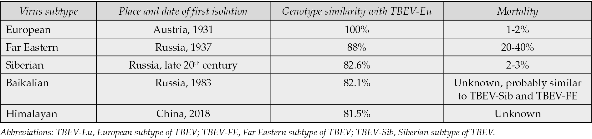
Traditionally, three main lineages have been recognized, European (TBEV-Eu), Siberian (TBEV-Sib), and Far Eastern (TBEV-FE), which differ in geographical distribution, genetic features, and virulence. Two additional subtypes, Baikalian and Himalayan, have been described more recently. The Baikalian subtype likely originated from a recombination between Siberian and Far-Eastern strains; the Himalayan subtype, identified in Marmota himalayana, shows distinct genetic features and can be divided into two lineages [11, 12].
Over time, genetic diversification has contributed to phenotypic variability, including differences in virulence and neurotropism [7]. Specific genomic elements, such as poly(A) regions, and amino acid substitutions in the E protein (particularly within domains II and III) have been implicated in modulating neuroinvasiveness and replication in the central nervous system [13]. Despite this variability, TBEV exhibits low substitution rate, and overall genetic divergence among subtypes remains limited compared to other flaviviruses, with amino-acid similarities between the strains ranging from 81% to 96%, with the European and the Himalayan subtypes differing the most from each other [7, 14].
Epidemiology and modes of transmission
TBEV is primarily transmitted through the bite of infected ticks, which constitute the main vector of the disease. Small mammals, particularly rodents and insectivores, serve as the principal reservoir hosts, sustaining viral circulation in natural foci. In contrast, large mammals, including humans, typically develop only transient and low-level viremia, insufficient to contribute meaningfully to onward transmission [8].
Ticks acquire the virus while feeding on infected hosts or, more efficiently, through co-feeding transmission, whereby infected and uninfected ticks feed in close proximity on the same host. This mechanism allows viral maintenance even in the absence of systemic viremia in the vertebrate host. Vertical transmission within ticks has also been described. Once infected, ticks can harbor the virus in their salivary glands throughout their lifespan, which may extend up to several years, enabling long-term persistence of TBEV in the environment [8].
Transmission to humans occurs during tick feeding, as virus-containing saliva is inoculated into the host. Notably, TBEV transmission may take place within minutes to a few hours after tick attachment, in contrast to other tick-borne pathogens that require prolonged feeding. The probability of transmission increases with feeding duration, although interrupted feeding followed by reattachment to a different host may further accelerate transmission [8]. Tick saliva itself plays an active role in facilitating infection, as it contains immunomodulatory factors that reduce host inflammatory responses and enhance viral dissemination [15].
In addition to vector-borne transmission, alimentary infection represents a well-recognized alternative route. Consumption of unpasteurized milk or dairy products from infected ruminants (particularly goats, sheep, and cows) can lead to infection and has been associated with localized outbreaks [16]. Other, less common transmission routes, including solid organ transplantation, blood transfusion, breastfeeding, and aerosol exposure, have been documented but remain rare [8]. Figure 1 shows the modes of transmission of TBE.
TBEV displays a heterogeneous geographic distribution that is primarily driven by the ecology of its primary vector, Ixodes ticks, and by environmental conditions that sustain enzootic transmission cycles (Figure 2).
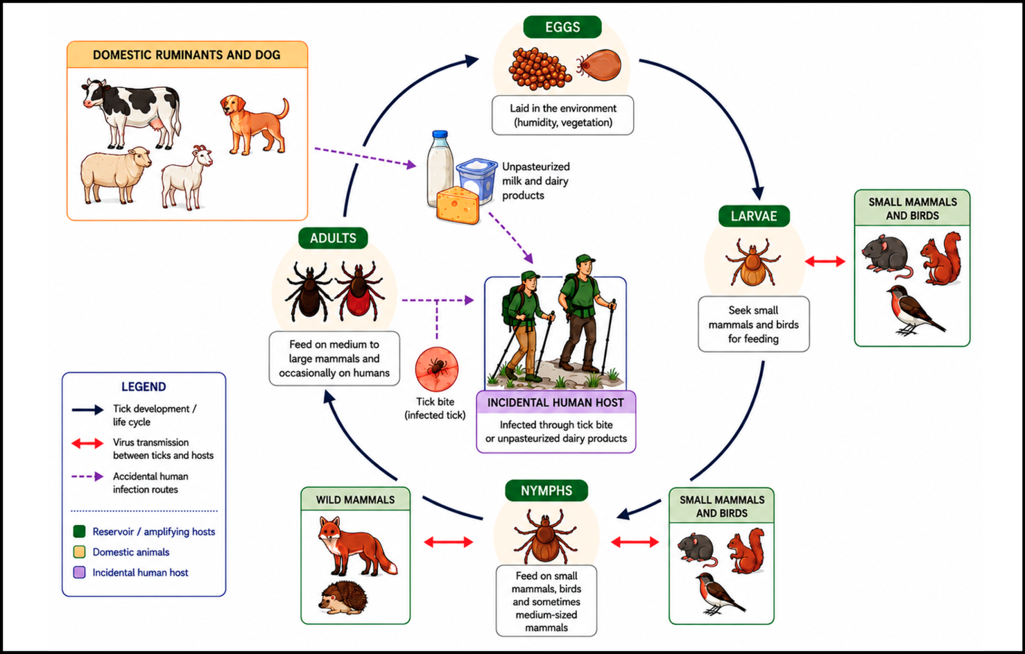
Figure 1 - TBE virus transmission cycle.
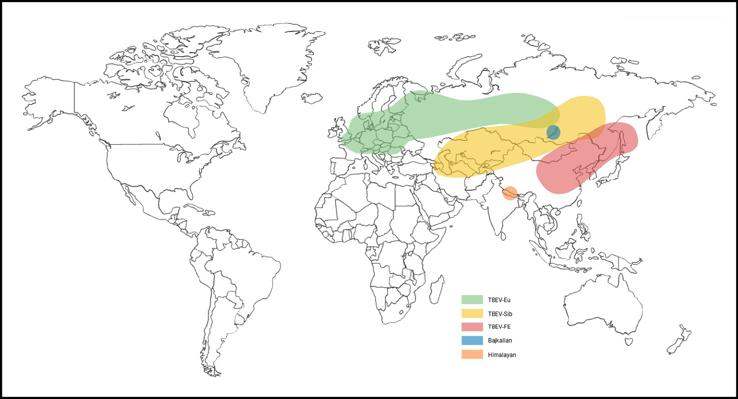
Figure 2 - Geographic distribution of TBE.
Across Europe and Asia, the virus is maintained in natural foci involving small mammals and ticks, with humans acting as incidental, dead-end hosts. The European subtype, transmitted mainly by Ixodes ricinus, predominates across Central and Western Europe, extending from the Baltic states and Scandinavia through Germany, Austria, Switzerland, and the Czech Republic to the Balkans and northern Italy. In contrast, the Siberian and Far Eastern subtypes, primarily associated with Ixodes persulcatus, circulate across Russia and large parts of Asia, including China, Japan, and the Korean peninsula. These lineages differ not only in geographic distribution but also in clinical severity, with higher case-fatality rates reported for infections caused by Far-Eastern strains (up to 20-40%, compared to 1-2% for TBEV-Eu) [2, 8].
From an epidemiological perspective, TBE represents one of the most relevant tick-borne viral infections in the Northern Hemisphere [1, 2]. The World Health Organization estimates that approximately 10,000-15,000 clinical cases are reported annually across the endemic regions. However, this figure likely underestimates the true burden of disease, as a substantial proportion of infections remain asymptomatic or present with non-specific clinical features and are therefore not diagnosed [1]. Incidence rates vary considerably both between and within countries, with the highest notification rates consistently observed in the Czech Republic, Lithuania, Latvia, Estonia, and Russia [17].
Over the past three decades, a progressive increase in TBE incidence has been documented. This trend is thought to result from a combination of ecological, climatic, and behavioural factors rather than improved surveillance alone [1, 3]. Climate change, in particular, has a profound impact on tick distribution and activity. Warmer temperatures, milder winters, and increased humidity have expanded the geographic range of Ixodes ticks and prolonged their seasonal activity [8, 18]. As a result, the transmission season, traditionally confined to spring and autumn, has lengthened, with tick activity increasingly documented during winter months in several European regions. In parallel, the expansion of ticks to higher altitudes and latitudes has introduced TBE risk into previously unaffected regions. Italy provides a paradigmatic example of the evolving epidemiology of TBE in Europe. Historically, autochthonous transmission was largely confined to the north-eastern regions, particularly Friuli-Venezia Giulia and Trentino-Alto Adige, which are ecologically contiguous with highly endemic areas of Central Europe [19, 20]. However, over the past two decades, new endemic foci have progressively emerged in previously unaffected regions, including parts of Veneto, Lombardy, and Piedmont, with sporadic evidence of transmission also reported in central Italy [1, 19]. This geographic expansion reflects broader European patterns and carries important public health implications, as it exposes populations with limited awareness and low vaccination coverage to an emerging infectious risk [20].
TBEV infection in humans typically begins at the site of a tick bite, where the virus is inoculated into the skin together with tick saliva during blood feeding [21]. After the tick attaches to the skin, kininase deactivation of bradykinin occurs, thereby reducing the pain related to the tick bite [22]. Anticoagulants, immunomodulators, and vasodilators, which are essential components of saliva composition, enhance viral entry and replication. Early infection occurs in the dermis, where TBEV infects neutrophils, monocytes, and skin-resident dendritic cells. These cells likely contribute to viral transport to regional lymph nodes [23]. At the same time, tick saliva exerts local immunomodulatory effects, attenuating macrophage recruitment and skewing the immune response toward a less effective Th2 profile [24]. Salivary components have also been shown to promote oxidative stress by increasing pro-oxidative molecules, such as 4-hydroxy-nonenal (4-HNE), while reducing antioxidant defenses. This imbalance contributes to cellular damage, sustains non-protective inflammation, and enhances TNF-α production [25].
Once TBEV enters cells, its pathogen-associated molecular patterns (PAMPs) are recognized by pattern recognition receptors (PRRs), including RIG-I–like receptors and Toll-like receptors (TLRs). Their activation triggers NF-κB and downstream pro-inflammatory cytokines, stimulating expression of interferon-stimulated genes (ISGs). These events enhance innate and adaptive immunity by enhancing antigen presentation, disrupting the viral life cycle, and activating macrophages, NK cells, and lymphocytes. The importance of this pathway is underscored by the observation that interferon response can reduce viral replication by several orders of magnitude [26]. Conversely, defects in early immune pathway, such as polymorphisms or reduced expression of TLR3 in dendritic or epithelial cells are linked to a higher risk of developing symptomatic or severe TBE [27].
Following initial amplification in skin immune cells, TBEV is transported to regional lymph nodes. Viral replication in draining lymph nodes is believed to generate viremia, enabling spread to peripheral tissues. Although the precise cell tropism in peripheral organs remains incompletely defined, dendritic cells, macrophages, and possibly neutrophils are thought to be major replication sites.
A critical determinant of disease severity is the ability of TBEV to invade the CNS. Neuroinvasion likely occurs during the viremic phase through multiple mechanisms, including transport via infected leukocytes (“Trojan horse” mechanism), direct infection of endothelial cells, and receptor-mediated entry into brain microvascular endothelial cells. Notably, experimental evidence suggests that disruption of the blood-brain-barrier (BBB) is not required for initial CNS entry, as BBB breakdown appears to occur later in the course of infection. TBEV can also access the CNS via retrograde axonal transport. Specific host receptors, such as low-density lipoprotein receptor-related protein 8, have been implicated in facilitating viral entry into neuronal cells.
Within the CNS, TBEV displays a marked tropism for neurons, where it replicates efficiently, reducing cell viability and spreading trans-synaptically. Infection of astrocytes and other glial cells has also been described in vitro [28, 29].
CNS damage in TBEV infection may result from direct neuronal injury or cell-cycle arrest in neural stem cell populations. However, immune-mediated mechanisms appear to play a predominant role, as infiltration of immune cells into the CNS contributes to neuronal loss and impaired connectivity. Studies evaluating inflammatory responses in serum and cerebrospinal fluid (CSF) have shown that, during the early phase of CNS involvement, innate and Th1 adaptive responses are predominantly detected in the CSF, whereas Th17 and B-cell responses are more prominent in the systemic circulation [30]. Such an increase in Th17 responses can contribute to CNS neutrophilic inflammation, which is further driven by the neutrophil chemoattractant IL-8 and amplified by TNF-αa. Similar findings have been described in conditions such as Lyme arthritis [31]. Moreover, activated cytotoxic T-cells can cross the BBB and contribute to TBEV-associated damage, characterized by neuronal loss, reactive gliosis, neuronophagia, and immune cell infiltration, which is observed mainly in the thalamus, cerebellum, caudate nucleus, and brainstem [9].
Biphasic disease course
TBE encompasses a broad and heterogeneous clinical spectrum, extending from asymptomatic or subclinical infection to severe neuroinvasive disease, possibly with long-term sequelae or fatal outcome. Approximately three-fourths of infections attributed to the European TBE virus subtype remain asymptomatic, while symptomatic cases typically manifest after an incubation period of 7-14 days (with a range of 2-28 days) after tick bite. In contrast, following consumption of unpasteurized milk or dairy products derived from infected sheep or goats, clinical symptoms usually develop in less than 2 weeks, often within 2-4 days [32].
When clinically evident, the illness classically follows a biphasic course, each with specific clinical manifestations. This distinctive temporal pattern is particularly associated with the European subtype virus (65-75% of patients), representing a hallmark of this infection. It consists of two distinct phases separated by a symptom-free interval. A monophasic disease course, characterized by isolated neurologic illness without a preceding febrile phase, is more commonly observed with Far Eastern and Siberian subtype infections and has been associated with more severe disease outcomes [8, 33]. The initial phase corresponds to systemic viremia and typically persists for 2-10 days (viremic prodromal phase). This early stage is characterized by a non-specific, flu-like syndrome, most commonly presenting with fever (99%), fatigue (63%), malaise (62%), headache, myalgia, and arthralgia (54%), reflecting a transient systemic inflammatory response [8, 33, 34]. Based on a case-series of 98 patients observed with prodromic symptoms of TBE, without evidence of neurologic involvement, the most frequent symptoms or signs were malaise and fatigue, fever, and headache, which typically resolve in 4-5 days. This phase may be associated with laboratory abnormalities such as leukopenia, thrombocytopenia, and elevated liver enzymes [35]. Following an initial phase, patients experience a symptom-free interval lasting approximately 7 days (range 1 to 21 days). This asymptomatic interval represents a critical diagnostic window, as TBE may not be suspected during the initial phase when symptoms are nonspecific. Approximately 20 to over 50% of infected individuals progress to a second disease phase characterized by the recurrence of fever and involvement of the CNS. As expected, most cases present with pleocytosis and high cerebrospinal fluid (CSF) proteins, and hyponatremia and high CSF protein levels were observed in those experiencing a severe course. Presentation without pleocytosis was reported in about 10% of the cases in a series from Norway [36]. The rate of symptom progression is highly variable among individuals [8, 33, 34, 37].
Neurological manifestations
The second phase of TBE encompasses a spectrum of neuroinvasive presentations whose clinical phenotype is largely determined by the specific CNS structures affected. The most frequent presentations are meningitis and meningoencephalitis. Clinically, the neurological symptoms present during this stage do not deviate significantly from those seen in typical acute viral meningoencephalitis. Meningitis, reported in roughly half of affected patients, typically manifests with fever, headache, vomiting, neck stiffness and photophobia. Meningoencephalitis, which occurs in approximately 40% of cases, is associated with more severe neurological impairment, including altered levels of consciousness, which is present in a third of cases and may range from somnolence to coma. In contrast with other viral meningoencephalitis, seizures are less common. Other manifestations can be tremor, ataxia, restlessness, muscle pain, dysarthria and fasciculations of the tongue [8, 34, 37]. The last one is a typical sign of TBE and should raise suspicion of diagnosis [38]. Paretic complications during the acute phase of the disease occur in about 10% of patients and are primarily due to spinal cord or peripheral nerve involvement. Bulbar symptoms are reported in fewer than 5% of cases and may progress to respiratory failure [39]. Less commonly, TBE infection may present as myelitis, observed in 5-10% of patients. In this case the clinical manifestations reflect the predilection of the virus for the anterior horn of the spinal cord. Respiratory muscle involvement can be present and may require mechanical ventilation. Rarely, the infection affects the medulla and the brainstem, posing the patient at risk of respiratory failure and severe complications associated with high mortality. Peripheral nervous system involvement has been reported, usually presenting as myeloradiculitis with severe pain in the back and limbs, weak muscle reflexes, and sensory disturbances [8, 34, 37]. Cranial nerves can be affected, most commonly presenting as peripheral unilateral facial palsy, but also affecting ocular, pharyngeal, vestibular, and auditory functions [33, 40]. The clinical spectrum of TBEV during pregnancy closely mirrors that of non-pregnant patients. Few case reports in the literature indicate no evidence of vertical transmission, with pregnancies culminating in normal outcomes and infants demonstrating typical development throughout their first year [41].
Neurological involvement typically worsens as patients age, characterized by isolated meningitis in children and more severe forms in the elderly population [33, 41, 42].
Functional sequelae are reported in about half of affected cases, with fatigue and motor deficits being the most frequently reported. Based on a study on 209 patients with laboratory-confirmed TBE, having severe symptoms during the acute phase and older age were stronger predictors of an unfavourable outcome [43].
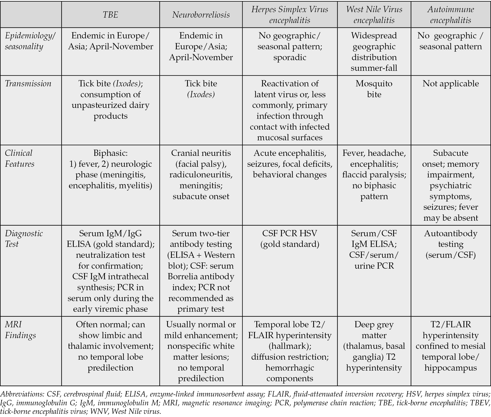
Table 2 shows the differential diagnoses of TBE.
Table 2 - Differential diagnosis of TBE.
DIAGNOSIS
TBE diagnosis is mainly based on clinical signs and symptoms, confirmed by serological and molecular tests on serum/blood and cerebrospinal fluid [2, 44]. According to the European Centre for Disease Prevention and Control (ECDC) Meeting Report in 2011, a confirmed TBE case requires both clinical symptoms of TBE and IgM plus IgG in serum or IgM in the CSF or IgM plus IgG in the CSF or detection of TBE viral nucleic acid in clinical specimen [45].
Following TBEV infection, in the initial phase of the disease, leukopenia and/or thrombocytopenia are often found, and liver function parameters may be altered. Leukocyte counts tend to improve over the course of illness, whereas thrombocytopenia and liver enzyme abnormalities may worsen [35, 46].
In the first viremic phase, which precedes seroconversion and the onset of central nervous system manifestations, TBE viral RNA can be detected in the patient’s blood or serum with specific molecular tests (NAT, Nucleic Acid Test), such as polymerase chain reaction (RT-PCR). Once neurological symptoms arise, however, TBEV RNA is hardly detected in the blood, although persistent viremia has been observed in immunocompromised individuals [19]. Viral RNA can be seldom detected in this second phase in the cerebrospinal fluid, if early collected, or in urine [47]. While RT-PCR in blood can be crucial for detecting TBEV in the initial phase of TBE, however it has few or none diagnostic value in the second phase of the infection, when most patients seek medical attention.
Confirmation of a suspected TBEV infection usually relies on detection of TBEV-specific IgM and IgG antibodies in serum. IgM antibodies can be detected in the blood from the onset of neurological symptoms (usually within 6 days of symptoms onset) up to several months after infection, while IgG antibodies persist lifelong and have a protective effect. Interindividual variations in antibody production and kinetics have been described.
Demonstration of intrathecal synthesis of anti-TBEV antibodies can also aid in confirming the diagnosis of neurological infection. Specific antibodies can be found in the cerebrospinal fluid in 50% of the patients early after onset of TBE disease, and by the 10-15th day of illness they almost invariably become detectable [44]. In the CSF, IgM production peaks later than in the blood, typically becoming detectable after 2 weeks [48].
The serological tests, mainly based on enzyme-linked immunosorbent assays (ELISA), generally demonstrate good sensitivity, but can pose interpretation problems due to flaviviruses extensive cross-reactivity and the so-called “original antigenic sin” phenomenon that occurs during sequential infections with other flaviviruses. In these cases, to avoid false-positive results, confirmatory neutralization tests are recommended, but these are performed only by specialized laboratories [44].
A previous vaccination with TBE can also be a confounding factor in the diagnosis of TBE. Current TBE vaccines available in Europe have demonstrated long-lasting immune responses, with neutralizing antibodies persisting for over 5 years. Patients who, despite vaccination, develop TBE (vaccine “breakthrough”) exhibit an immunological response characterized by delayed development of the specific IgM response, along with rapid increase in serum IgG antibodies [49]. Unfortunately, the currently available standard diagnostic tests do not allow for a differential diagnosis between vaccination-induced immunity and infection-induced immunity but IgG avidity testing can be of some help [50]. Systems that would allow for the discrimination of IgG and IgM responses to the viral protein NS1, which is present in in vivo infections but not in the inactivated virus vaccine preparations available in Europe, are recently being studied [51].
TREATMENT
Despite its potentially severe and life-threatening manifestations, there is currently no proven or approved, specific antiviral therapy for TBE. Clinical management is therefore primarily supportive and guided by disease severity. For neuroinvasive disease requiring hospitalization, supportive care includes adequate fluid and electrolyte management, along with antipyretics and antiemetics. Moreover, anticonvulsant treatment can be started in presence of seizures. In parallel, careful neurological assessment and close clinical monitoring are essential to detect early signs of deterioration. This approach allows timely escalation of care, including admission to an intensive care unit (ICU) when indicated, where airway protection, mechanical ventilation, and hemodynamic support can be promptly initiated [8, 52].
Among the neurological complications that can aggravate the clinical picture and worsen the outcomes, cerebral edema, bulbar dysfunction and neuromuscular paralysis are the most threatening. Regarding cerebral edema, the use of mannitol and/or corticosteroids is recommended in order to reduce intracranial pressure [53].
Although corticosteroids are recommended in certain bacterial meningitides and have been proposed as a potential immunomodulatory treatment in severe cases of TBE, their role remains controversial. In endemic areas, they are often used in severe cases based on anecdotal clinical experience [54]. However, observational studies show no clear clinical benefit, and treated patients have sometimes experienced longer hospitalization, although corticosteroid use was not independently associated with incomplete recovery [55]. Overall, the evidence is inconsistent and largely retrospective, underscoring the need for randomized controlled trials. A multicentre double-blind study of dexamethasone in adults with TBE has been proposed to assess whether dexamethasone improves neuropsychological and quality-of-life outcomes in patients with TBE and central nervous system involvement [4]. At present, corticosteroids are not standard therapy and should be considered on a case-by-case basis.
Immunotherapy-based strategies have been reported, with some cases describing favorable outcomes.
As in other flaviviral infections, high-dose intravenous immunoglobulin (IVIG) has been proposed for the treatment of TBE [56]. However, its use remains controversial due to the potential risk of antibody-dependent enhancement (ADE), a phenomenon in which sub-neutralizing antibodies facilitate viral entry into Fc receptor-bearing immune cells, potentially increasing viral replication and disease severity [8].
In experimental studies, the TBEV-specific antibody content of some IVIG batches showed strong neutralizing activity in cell culture and was associated with protection in mice [57]. Nevertheless, IVIG alone may be insufficient in immunodeficient patients with severe disease. In such cases, the combination of plasma transfusions containing TBEV-IgG and corticosteroids has been proposed to increase neutralizing antibody titres and potentially support viral clearance [58], although evidence is currently limited to case-based reports.
There is growing interest in whether nucleoside and nucleotide analogs (NAs), mostly drugs repurposed from other infections, could have a role in TBE, but evidence base remains largely preclinical, with only isolated human observations [59].
Favipiravir, a broad-spectrum antiviral, has generated particular interest following a published off-label case report in a patient with severe TBE who subsequently recovered [60]. Acting as a pro-drug, it is recognized by the RNA-dependent RNA polymerase as a pseudopurine and it could inhibit viral replication, as demonstrated for West Nile virus (WNV) [61].
Other NAs, including remdesivir and sofosbuvir, have shown in vitro inhibitory activity against TBEV, and have raised early signals related to resistance selection, which supports further evaluation rather than clinical use [62].
More recently, a preprint found that bemnifosbuvir, either by itself or paired with remdesivir, reduced viral replication in ex vivo and organoid-like models, suggesting potential activity but remaining strictly preclinical [63].
Finally, ribavirin can inhibit TBEV replication in cell culture and protect infected cells from cytopathic effect, yet its clinical value in TBE remains unproven, and reviews generally conclude that interferon/ribavirin-based approaches have not produced convincing benefit [64].
Overall, these antivirals remain investigational, and controlled clinical studies would be required before any could be recommended beyond compassionate/off-label use in selected cases.
These direct antiviral agents (DAAs), together with TBEV monoclonal antibodies, have been recognized as the most promising pipelines for TBE treatment. As for SARS-CoV-2 and respiratory syncytial virus, monoclonal antibodies (mAb) are already an established strategy for the prevention and treatment of severe viral infections [65,66].
Treatment with TBEV-specific monoclonal antibodies has already demonstrated efficacy in mouse models [67]. Building on this rationale, and on the clinical need for options in severe TBE, particularly in immunocompromised patients with suspected ongoing viral replication, TBE therapeutics are now starting to move beyond preclinical work. In fact, a first-in-human trial of an investigational TBEV monoclonal antibody (e.g. TBE025) is listed in trial registries and is enrolling healthy adults to assess safety, tolerability, and pharmacokinetics and to define a recommended Phase II dose [68]. While this is a clear milestone for the field, it remains an early step, and clinical efficacy in patients will need to be demonstrated in subsequent studies.
VACCINES AND PREVENTION
Available TBE vaccines
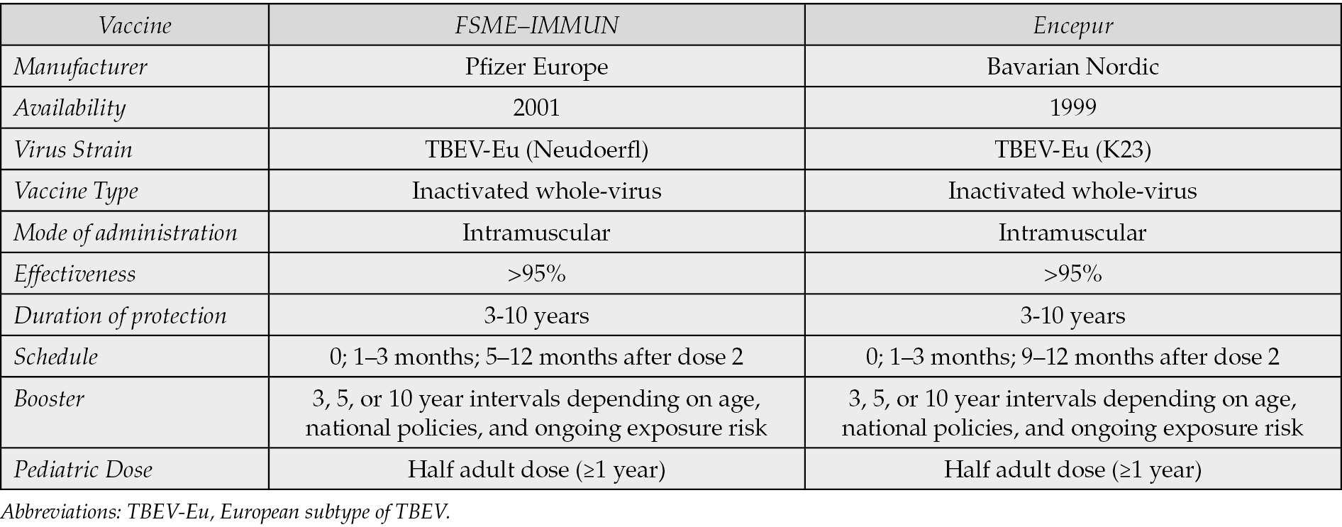
At present, TBE can be effectively prevented through active immunization [69]. Two vaccines are licensed in Europe: FSME-IMMUN (Pfizer Europe, Brussels, Belgium), derived from a 1979 formulation and available since 2001; and Encepur (Bavarian Nordic, Hellerup, Denmark), licensed in Germany in 1991 and available across Europe since 1999 [70]. Table 3 shows their main characteristics. Both products include pediatric formulations containing half the adult dose and are authorized for children aged ≥1 year [71].
The two vaccines are inactivated whole-virus preparations based on European TBEV strains (Neudoerfl for FSME–IMMUN and K23 for Encepur). Vaccine production involves propagation of TBE virus in chick embryo fibroblast cells, formaldehyde inactivation, and purification by ultracentrifugation. The virions are adsorbed onto aluminum hydroxide and stabilized with human serum albumin; both formulations are preservative-free and contain no thiomersal [33, 34].
Table 3 - Available vaccines in Europe and vaccination schedules.
Vaccination schedules and target populations
The standard primary immunization schedule comprises three intramuscular doses: the second 1–3 months after the first, and the third 5–12 months after the second. Completion of the primary series is recommended at least one week prior to potential exposure [33]. Accelerated schedules with shortened intervals between doses may be considered under particular circumstances when rapid protection is required [72, 73]. A booster dose may be administered no earlier than three years after the primary series, with subsequent boosters scheduled at 3-, 5-, or 10-year intervals depending on age, national policies, and ongoing exposure risk [33, 69].
Vaccination is recommended for all individuals aged ≥1 year living in highly endemic regions (≥5 cases/100,000/year), according to WHO and European Academy of Neurology guidelines [2]. In areas of lower incidence, immunization is targeted to at-risk populations, including individuals with occupational or recreational outdoor exposure, such as forest workers and hikers. Travelers to endemic areas with planned open-air activity, particularly during peak tick season in summer, should also be vaccinated [33]. A recent review of European policies found that nationwide programs are implemented only in seven countries; elsewhere, recommendations are limited to travellers or residents in endemic areas, with endemicity defined variably by incidence (e.g., ≥15 cases/100,000/year in Finland) or geography (e.g., below 1400 m in Italy) [20].
Immunogenicity, effectiveness, and safety
Evidence from the literature indicates that both licensed TBE vaccines induce near-complete seroconversion in adults and children following the three-dose primary series [17]. Antibody titers remain high in at least 95% of vaccinated individuals for up to three years, and two European studies reported detectable anti–TBEV neutralizing antibodies up to 10 years after a booster [74, 75]. Seropositivity is lower in immunosuppressed individuals [76] and gradually declines in those >60 years [77].
With the exception of one pediatric study [78], current findings support the interchangeability of the two vaccines for primary and booster immunization, with preserved immunogenicity [79]. Moreover, the neutralizing antibodies elicited confer cross-protection against the three main TBEV subtypes, as demonstrated in preclinical and human studies [17].
Randomized controlled trials were not conducted during vaccine approval; however post-licensure observational studies and systematic reviews demonstrate 90–99% effectiveness for both formulations in preventing clinical disease across all age groups, with consequent reductions in hospitalizations and mortality [80,81]. Breakthrough infections are rare and primarily affect older adults, suggesting booster intervals may need shortening in this population [77].
More than 75 million TBE vaccine doses administered in Europe since 2001 indicate a favorable safety profile [33]. Adverse events are more frequent following primary series doses [82] and in young children [83]. Serious reactions are exceedingly rare [83, 84], with most events being mild to moderate - commonly injection site pain, headache, fatigue, myalgia, or fever - and resolving spontaneously [77].
Preventive strategies beyond vaccination
Since vaccination coverage and adherence can be suboptimal [85], complementary preventive measures are essential. Tick control relies mainly on ectoparasiticides, though resistance may develop. Personal protection includes avoiding areas with dense foliage, wearing long pants that cover the ankles, avoiding tall grass or brush and avoiding unpasteurized dairy products [18, 32, 85]. After potential exposure, a thorough skin check and shower are recommended to remove unattached ticks, while attached ticks should be removed promptly by pulling straight out with tweezers or fingers [86].
Alongside vaccination, a One Health approach can provide a consistent pillar of TBE prevention. Moreover, through integrated surveillance and monitoring of disease drivers and early warning signals, One Health can help mitigate spillover events [87].
TBE virus circulates primarily in enzootic cycles involving Ixodes ticks and vertebrate hosts, with small mammals acting as key reservoirs and larger mammals (especially cervids) sustaining tick populations by providing blood meals and, through their abundance and movements patterns, shaping tick density and distribution across landscapes [88].
As with other vector-borne diseases (VBDs), tick-borne diseases (TBDs) are highly climate-sensitive because ticks are poikilotherms, and environmental temperature strongly influences core physiological processes such as blood-meal digestion, development, oviposition, and refeeding [89].
In addition, climate changes with warming temperatures, altered precipitation, and shorter winters may extend seasonal tick activity, facilitate expansion to higher altitudes and latitudes, and shift host distributions [90].
Comparable patterns have already been observed for other VBDs in Europe. For example, WNV seasons have intensified in recent years, and attribution modelling supports climate change as a driver of the long-term expansion of ecologically suitable area for WNV circulation in Europe [91,92]. Beyond climate, anthropogenic land-use change (e.g., reforestation, woodland fragmentation, urban–forest interfaces, agricultural abandonment) can increase edge habitats, intensifying and favoring host–tick encounters.
A One Health framework therefore prioritizes integrated surveillance that links human case reporting with veterinary and wildlife indicators, entomological monitoring (tick density, infection prevalence), and environmental data (climate, vegetation, landscape structure). This approach enables early detection of outbreaks, risk mapping, and seasonal forecasting [93]. Importantly, One Health is a governance model that aligns ministries, public health agencies, forestry and wildlife services, veterinarians, and communities around shared metrics, transparent data sharing, and adaptive interventions [94]. For research, it encourages cross-disciplinary study of how host community composition, biodiversity, and anthropogenic pressures modulate virus persistence; how microclimate and habitat management influence tick phenology; and how human mobility and vaccine uptake shape outbreak dynamics.
CONCLUSIONS
TBE is an emerging infection with increasing relevance in endemic areas. Although most cases are mild, severe neurological disease can occur. In the absence of specific treatment, vaccination and preventive measures remain essential. A One Health approach, integrating human, animal, and environmental surveillance, together with increased awareness, is important to limit the impact of the disease.
Conflict of interest
Authors have no conflict of interest to disclose.
Funding
Authors received no funding for this study.
References
[1] Riccardi N, Antonello RM, Luzzati R, Zajkowska J, Di Bella S, Giacobbe DR. Tick-borne encephalitis in Europe: a brief update on epidemiology, diagnosis, prevention, and treatment. Eur J Intern Med. 2019; 62: 1-6. doi: 10.1016/j.ejim.2019.01.004.
[2] Taba P, Schmutzhard E, Forsberg P, et al. EAN consensus review on prevention, diagnosis and management of tick-borne encephalitis. Eur J Neurol. 2017; 24(10): 1214-e61. doi: 10.1111/ene.13356.
[3] Beerlage-de Jong N, Blanford J. Mapping the risk of tick-borne encephalitis in Europe for informed vaccination decisions. J Travel Med. 2025; 32(2). doi: 10.1093/jtm/taae153.
[4] Stupica D, Kejžar N, Collinet-Adler S, Bajrović FF. Which trial do we need? Dexamethasone therapy in adults with tick-borne encephalitis: a double-blind placebo-controlled multicentre randomized trial. Clin Microbiol Infect. 2024; 30(11): 1344-1346. doi: 10.1016/j.cmi.2024.06.004.
[5] Deviatkin AA, Aleshina YA, Karganova GG, Lukashev AN. Selection Pressure Profile Suggests Species Criteria among Tick-Borne Orthoflaviviruses. Viruses. 2024; 16(10). doi: 10.3390/v16101554.
[6] Simmonds P, Butković A, Grove J, et al. Taxonomic expansion and reorganization of Flaviviridae. Nat Microbiol. 2025; 10(11): 3026-3037. doi: 10.1038/s41564-025-02134-0.
[7] Bondaryuk AN, Kulakova NV, Belykh OI, Bukin YS. Dates and Rates of Tick-Borne Encephalitis Virus-The Slowest Changing Tick-Borne Flavivirus. Int J Mol Sci. 2023; 24(3). doi: 10.3390/ijms24032921.
[8] Pustijanac E, Buršić M, Talapko J, Škrlec I, Meštrović T, Lišnjić D. Tick-Borne Encephalitis Virus: A Comprehensive Review of Transmission, Pathogenesis, Epidemiology, Clinical Manifestations, Diagnosis, and Prevention. Microorganisms. 2023; 11(7). doi: 10.3390/microorganisms11071634.
[9] Worku DA. Tick-Borne Encephalitis (TBE): From Tick to Pathology. J Clin Med. 2023; 12(21). doi: 10.3390/jcm12216859.
[10] Belikov SI, Kondratov IG, Potapova UV, Leonova GN. The relationship between the structure of the tick-borne encephalitis virus strains and their pathogenic properties. PLoS One. 2014; 9(4): e94946. doi: 10.1371/journal.pone.0094946.
[11] Sukhorukov GA, Paramonov AI, Lisak OV, et al. The Baikal subtype of tick-borne encephalitis virus is evident of recombination between Siberian and Far-Eastern subtypes. PLoS Negl Trop Dis. 2023; 17(3): e0011141. doi: 10.1371/journal.pntd.0011141.
[12] Dai X, Shang G, Lu S, Yang J, Xu J. A new subtype of eastern tick-borne encephalitis virus discovered in Qinghai-Tibet Plateau, China. Emerg Microbes Infect. 2018; 7(1): 74. doi: 10.1038/s41426-018-0081-6.
[13] Asghar N, Lee YP, Nilsson E, et al. The role of the poly(A) tract in the replication and virulence of tick-borne encephalitis virus. Sci Rep. 2016; 6: 39265. doi: 10.1038/srep39265.
[14] Wang R, Gu A, Li F, et al. Evolutionary Patterns and Genotype-Specific Amino Acid Mutations of Tick-Borne Encephalitis Virus. Int J Mol Sci. 2025; 26(3). doi: 10.3390/ijms26030954.
[15] Hart CE, Ribeiro JM, Kazimirova M, Thangamani S. Tick-Borne Encephalitis Virus Infection Alters the Sialome of Ticks During the Earliest Stages of Feeding. Front Cell Infect Microbiol. 2020; 10: 41. doi: 10.3389/fcimb.2020.00041.
[16] Ličková M, Fumačová Havlíková S, Sláviková M, Klempa B. Alimentary Infections by Tick-Borne Encephalitis Virus. Viruses. 2021; 14(1). doi: 10.3390/v14010056.
[17] Amicizia D, Domnich A, Panatto D, et al. Epidemiology of tick-borne encephalitis (TBE) in Europe and its prevention by available vaccines. Hum Vaccin Immunother. 2013; 9(5): 1163-1171. doi: 10.4161/hv.23802.
[18] Kwasnik M, Rola J, Rozek W. Tick-Borne Encephalitis-Review of the Current Status. J Clin Med. 2023; 12(20). doi: 10.3390/jcm12206603.
[19] Caracciolo I, Bassetti M, Paladini G, et al. Persistent viremia and urine shedding of tick-borne encephalitis virus in an infected immunosuppressed patient from a new epidemic cluster in North-Eastern Italy. J Clin Virol. 2015; 69: 48-51. doi: 10.1016/j.jcv.2015.05.019.
[20] Srinivasan R, Angulo FJ, Duench S, et al. Tick-borne encephalitis in international travellers: a systematic review and vaccine recommendations. J Travel Med. 2025; 32(8). doi: 10.1093/jtm/taaf115.
[21] Suppan J, Engel B, Marchetti-Deschmann M, Nürnberger S. Tick attachment cement - reviewing the mysteries of a biological skin plug system. Biol Rev Camb Philos Soc. 2018; 93(2): 1056-1076. doi: 10.1111/brv.12384.
[22] Mulenga A, Kim T, Ibelli AMG. Amblyomma americanum tick saliva serine protease inhibitor 6 is a cross-class inhibitor of serine proteases and papain-like cysteine proteases that delays plasma clotting and inhibits platelet aggregation. Insect Mol Biol. 2013; 22(3): 306-319. doi: 10.1111/imb.12024.
[23] Wang B, Papdi N. Deep Subcutaneous Tick Embedding Following Prolonged Attachment: A Case Report and Mini-Review of Tick Fixation Mechanisms. Cureus. 2026; 18(1): e102067. doi: 10.7759/cureus.102067.
[24] Schneider CA, Calvo E, Peterson KE. Arboviruses: How Saliva Impacts the Journey from Vector to Host. Int J Mol Sci. 2021; 22(17). doi: 10.3390/ijms22179173.
[25] Dobrzyńska M, Moniuszko-Malinowska A, Jarocka-Karpowicz I, Czupryna P, Groth M, Skrzydlewska E. Metabolic Response to Tick-Borne Encephalitis Virus Infection and Bacterial Co-Infections. Pathogens. 2022; 11(4). doi: 10.3390/pathogens11040384.
[26] Sui L, Wang W, Guo X, et al. Multi-protomics analysis identified host cellular pathways perturbed by tick-borne encephalitis virus infection. Nat Commun. 2024; 15(1): 10435. doi: 10.1038/s41467-024-54628-w.
[27] Fortova A, Barkhash AV, Pychova M, et al. Genetic polymorphisms in innate immunity genes influence predisposition to tick-borne encephalitis. J Neurovirol. 2023; 29(6): 699-705. doi: 10.1007/s13365-023-01182-8.
[28] Yao YL, Chen J, Si H, Shi Y, Luo R. LRP8-dependent neurotropism of tick-borne encephalitis virus: From species-specific entry gateway to zoonotic spillover and receptor-mimetic therapy. Innovation (Camb). 2026; 7(3): 101226. doi: 10.1016/j.xinn.2025.101226.
[29] Li P, Hui S, Chong Z, et al. LRP8 is an entry receptor for tick-borne encephalitis viruses. Proc Natl Acad Sci U S A. 2025; 122(44): e2525771122. doi: 10.1073/pnas.2525771122.
[30] Bogovič P, Lusa L, Korva M, et al. Inflammatory Immune Responses in the Pathogenesis of Tick-Borne Encephalitis. J Clin Med. 2019; 8(5). doi: 10.3390/jcm8050731.
[31] Strle K, Sulka KB, Pianta A, et al. T-Helper 17 Cell Cytokine Responses in Lyme Disease Correlate With Borrelia burgdorferi Antibodies During Early Infection and With Autoantibodies Late in the Illness in Patients With Antibiotic-Refractory Lyme Arthritis. Clin Infect Dis. 2017; 64(7): 930-938. doi: 10.1093/cid/cix002.
[32] Centers for Disease Control and Prevention (CDC). Tick-borne encephalitis. Atlanta (GA): CDC; 2025. Available at: https://www.cdc.gov/yellow-book/hcp/travel-associated-infections-diseases/tick-borne-encephalitis.html. [accessed 22 Feb 2026].
[33] Hills SL. Tick-borne encephalitis vaccine: recommendations of the Advisory Committee on Immunization Practices, United States, 2023. MMWR Recomm Rep. 2023; 72(5): 1-28. Available at: https://www.cdc.gov/mmwr/volumes/72/rr/rr7205a1.htm. [accessed 22 Feb 2026].
[34] Lindquist L, Vapalahti O. Tick-borne encephalitis. Lancet. 2008; 371(9627): 1861-1871. doi: 10.1016/S0140-6736(08)60800-4.
[35] Bogovič P, Kastrin A, Lotrič-Furlan S, et al. Clinical and Laboratory Characteristics and Outcome of Illness Caused by Tick-Borne Encephalitis Virus without Central Nervous System Involvement. Emerg Infect Dis. 2022; 28(2): 291-301. doi: 10.3201/eid2802.211661.
[36] Skudal H, Lorentzen ÅR, Stenstad T, et al. Clinical characteristics and factors affecting disease severity in hospitalized tick-borne encephalitis patients in Norway from 2018 to 2022. Eur J Clin Microbiol Infect Dis. 2024; 43(7): 1355-1366. doi: 10.1007/s10096-024-04855-2.
[37] Chiffi G, Grandgirard D, Leib SL, Chrdle A, Růžek D. Tick-borne encephalitis: A comprehensive review of the epidemiology, virology, and clinical picture. Rev Med Virol. 2023; 33(5): e2470. doi: 10.1002/rmv.2470.
[38] Benvenuto N, Sartori A, Di Bella S. Tongue tremor suggesting tick-borne encephalitis. Pract Neurol. 2025; 25(5): 485-486. doi: 10.1136/pn-2024-004434.
[39] Smíšková D, Pícha D, Slížek M, Džupová O. Paretic complications of tick-borne encephalitis and Lyme neuroborreliosis in the Czech Republic: Characteristics and clinical outcome. Ticks Tick Borne Dis. 2024; 15(2): 102302. doi: 10.1016/j.ttbdis.2023.102302.
[40] Lotric-Furlan S, Strle F. Peripheral facial palsy in patients with tick-borne encephalitis. Clin Microbiol Infect. 2012; 18(10): 1027-1032. doi: 10.1111/j.1469-0691.2011.03719.x.
[41] Bjonholm E, Soderholm S, Stephansson O, Askling HH. Tick-borne encephalitis in pregnant women: A mini narrative review. New Microbes New Infect. 2022; 48: 101017. doi: 10.1016/j.nmni.2022.101017.
[42] Kohlmaier B, Schweintzger NA, Sagmeister MG, et al. Clinical Characteristics of Patients with Tick-Borne Encephalitis (TBE): A European Multicentre Study from 2010 to 2017. Microorganisms. 2021; 9(7). doi: 10.3390/microorganisms9071420.
[43] Slížek M, Chovanec L, Pražák V, et al. Complications and long-term sequelae of tick-borne encephalitis: association with age and other predictors - a multicenter observational study. Ticks Tick Borne Dis. 2026; 17(3): 102624. doi: 10.1016/j.ttbdis.2026.102624.
[44] Holzmann H. Diagnosis of tick-borne encephalitis. Vaccine. 2003; 21 Suppl. 1: S36-S40. doi: 10.1016/s0264-410x(02)00819-8.
[45] European Centre for Disease Prevention and Control. Tick-borne diseases: meeting report. Stockholm: ECDC; 2011. Available at: https://www.ecdc.europa.eu/sites/default/files/media/en/publications/Publications/Tick-borne-diseases-meeting-report.pdf. [accessed 1 May 2026].
[46] Lotric-Furlan S, Strle F. Thrombocytopenia, leukopenia and abnormal liver function tests in the initial phase of tick-borne encephalitis. Zentralbl Bakteriol. 1995; 282(3): 275-278. doi: 10.1016/s0934-8840(11)80127-1. PMID: 7549159.
[47] Veje M, Studahl M, Norberg P, et al. Detection of tick-borne encephalitis virus RNA in urine. J Clin Microbiol. 2014; 52(11): 4111-4112. doi: 10.1128/JCM.02428-14.
[48] Günther G, Haglund M, Lindquist L, Sköldenberg B, Forsgren M. Intrathecal IgM, IgA and IgG antibody response in tick-borne encephalitis. Long-term follow-up related to clinical course and outcome. Clin Diagn Virol. 1997; 8(1): 17-29. doi: 10.1016/s0928-0197(97)00273-0.
[49] Remoli ME, Marchi A, Fortuna C, et al. Anti-tick-borne encephalitis (TBE) virus neutralizing antibodies dynamics in natural infections versus vaccination. Pathog Dis. 2015; 73(2): 1-3. doi: 10.1093/femspd/ftu002.
[50] Vilibic-Cavlek T, Barbic L, Stevanovic V, Petrovic G, Mlinaric-Galinovic G. IgG Avidity: an Important Serologic Marker for the Diagnosis of Tick-Borne Encephalitis Virus Infection. Pol J Microbiol. 2016; 65(1): 119-121. Available at: https://reference-global.com/article/10.5604/17331331.1197285?tab=abstract. [accessed 1 May 2026].
[51] Girl P, Bestehorn-Willmann M, Zange S, Borde JP, Dobler G, von Buttlar H. Tick-Borne Encephalitis Virus Nonstructural Protein 1 IgG Enzyme-Linked Immunosorbent Assay for Differentiating Infection versus Vaccination Antibody Responses. J Clin Microbiol. 2020; 58(4). doi: 10.1128/JCM.01783-19.
[52] Bogovic P, Strle F. Tick-borne encephalitis: A review of epidemiology, clinical characteristics, and management. World J Clin Cases. 2015; 3(5): 430-441. doi: 10.12998/wjcc.v3.i5.430.
[53] Kumar G, Kalita J, Misra UK. Raised intracranial pressure in acute viral encephalitis. Clin Neurol Neurosurg. 2009; 111(5). Available at: https://pubmed.ncbi.nlm.nih.gov/19372001/. [accessed 1 May 2026].
[54] By JA, Dos AHH, Stenor C. Exploring corticosteroid therapy in tick-borne encephalitis: A case report and rationale for future research. Diagn Microbiol Infect Dis. 2025; 113(3): 116964. doi: 10.1016/j.diagmicrobio.2025.116964.
[55] Krawczuk K, Czupryna P, Pancewicz S, Ołdak E, Moniuszko-Malinowska A. Comparison of tick-borne encephalitis between children and adults-analysis of 669 patients. J Neurovirol. 2020; 26(4): 565-571. doi: 10.1007/s13365-020-00856-x.
[56] Růžek D, Dobler G, Niller HH. May early intervention with high dose intravenous immunoglobulin pose a potentially successful treatment for severe cases of tick-borne encephalitis? BMC Infect Dis. 2013; 13: 306. doi: 10.1186/1471-2334-13-306.
[57] Elsterova J, Palus M, Sirmarova J, Kopecky J, Niller HH, Ruzek D. Tick-borne encephalitis virus neutralization by high dose intravenous immunoglobulin. Ticks Tick Borne Dis. 2017; 8(2): 253-258. doi: 10.1016/j.ttbdis.2016.11.007.
[58] Hedin W, Bergman P, Akhirunessa M, et al. Severe Tick-Borne Encephalitis (TBE) in a Patient with X-Linked Agammaglobulinemia; Treatment with TBE Virus IgG Positive Plasma, Clinical Outcome and T Cell Responses. J Clin Immunol. 2024; 44(5): 116. doi: 10.1007/s10875-024-01718-5.
[59] Gejji V, Svoboda P, Stefanik M, et al. An RNA-dependent RNA polymerase inhibitor for tick-borne encephalitis virus. Virology. 2020; 546: 13-19. doi: 10.1016/j.virol.2020.03.006.
[60] Bologheanu R, Schubert L, Thurnher M, et al. Unexpected complete recovery of a patient with severe tick-borne encephalitis treated with favipiravir. Antiviral Res. 2020; 184. Available at: https://pubmed.ncbi.nlm.nih.gov/33058928/. [accessed 1 May 2026].
[61] Escribano-Romero E, de Oya N J, Domingo E, Saiz JC. Extinction of West Nile Virus by Favipiravir through Lethal Mutagenesis. Antimicrob Agents Chemother. 2017; 61(11). Available at: https://pubmed.ncbi.nlm.nih.gov/28848019/. [accessed 1 May 2026].
[62] Nyström K, Trybala E, Said J, et al. Remdesivir is active against tick-borne encephalitis virus and selects for resistance mutations in the viral RNA-dependent RNA polymerase. Infect Dis (Lond). 2025; 57(7): 628-635. doi: 10.1080/23744235.2025.2468510.
[63] Leoni S, Schultz-Pernice I, Krátka Z, et al. Bemnifosbuvir and remdesivir inhibit tick-borne encephalitis virus infection in complementary in vitro and ex vivo disease models. bioRxiv. 2025. doi: 10.1101/2025.11.13.688315.
[64] Tang WD, Tang HL, Peng HR, Ren RW, Zhao P, Zhao LJ. Inhibition of tick-borne encephalitis virus in cell cultures by ribavirin. Front Microbiol. 2023; 14: 1182798. Available at: https://www.frontiersin.org/journals/microbiology/articles/10.3389/fmicb.2023.1182798/pdf. [accessed 1 May 2026].
[65] Stadler E, Burgess MT, Schlub TE, et al. Monoclonal antibody levels and protection from COVID-19. Nat Commun. 2023; 14(1): 4545. doi: 10.1038/s41467-023-40204-1.
[66] Drysdale SB, Cathie K, Flamein F, et al. Nirsevimab for Prevention of Hospitalizations Due to RSV in Infants. N Engl J Med. 2023. Available at: https://www.nejm.org/doi/full/10.1056/NEJMoa2309189. [accessed 1 May 2026].
[67] Agudelo M, Palus M, Keeffe JR, et al. Broad and potent neutralizing human antibodies to tick-borne flaviviruses protect mice from disease. J Exp Med. 2021; 218(5): e20210236. Available at: https://pmc.ncbi.nlm.nih.gov/articles/PMC8040517/. [accessed 1 May 2026].
[68] ClinicalTrials.gov. First-in-human study testing a new antibody treatment for tick-borne encephalitis in healthy volunteers (SVRI01). 2025. Available at: https://clinicaltrials.gov/study/NCT07377253. [accessed 1 May 2026].
[69] World Health Organization. Vaccines against tick-borne encephalitis: WHO position paper—recommendations. Vaccine. 2011; 29(48): 8769-8770. doi: 10.1016/j.vaccine.2011.07.024.
[70] Kollaritsch H, Paulke-Korinek M, Holzmann H, Hombach J, Bjorvatn B, Barrett A. Vaccines and vaccination against tick-borne encephalitis. Expert Rev Vaccines. 2012; 11(9): 1103-1119. doi: 10.1586/erv.12.86.
[71] Girgsdies OE, Rosenkranz G. Tick-borne encephalitis: development of a paediatric vaccine. A controlled, randomized, double-blind and multicentre study. Vaccine. 1996; 14(15): 1421-1428. doi: 10.1016/s0264-410x(96)00081-3.
[72] Galgani I, Bunge EM, Hendriks L, Schludermann C, Marano C, De Moerlooze L. Systematic literature review comparing rapid 3-dose administration of the GSK tick-borne encephalitis vaccine with other primary immunization schedules. Expert Rev Vaccines. 2017; 16(9): 919-932. doi: 10.1080/14760584.2017.1358620.
[73] Zens K, Torgler R, Horn M, Larsen CS. Fast-Track to Protection? A Review of Encepur’s Express Dosing Schedule for Tick-Borne Encephalitis. Viruses. 2025; 17(11). doi: 10.3390/v17111439.
[74] Beran J, Lattanzi M, Xie F, Moraschini L, Galgani I. Second five-year follow-up after a booster vaccination against tick-borne encephalitis following different primary vaccination schedules demonstrates at least 10 years antibody persistence. Vaccine. 2019; 37(32): 4623-4629. doi: 10.1016/j.vaccine.2017.12.081.
[75] Paulke-Korinek M, Kundi M, Laaber B, et al. Factors associated with seroimmunity against tick borne encephalitis virus 10 years after booster vaccination. Vaccine. 2013; 31(9): 1293-1297. doi: 10.1016/j.vaccine.2012.12.075.
[76] Hertzell KB, Pauksens K, Rombo L, Knight A, Vene S, Askling HH. Tick-borne encephalitis (TBE) vaccine to medically immunosuppressed patients with rheumatoid arthritis: A prospective, open-label, multi-centre study. Vaccine. 2016; 34(5): 650-655. doi: 10.1016/j.vaccine.2015.12.029.
[77] Rampa JE, Askling HH, Lang P, et al. Immunogenicity and safety of the tick-borne encephalitis vaccination (2009-2019): A systematic review. Travel Med Infect Dis. 2020; 37: 101876. doi: 10.1016/j.tmaid.2020.101876.
[78] Wittermann C, Schöndorf I, Gniel D. Antibody response following administration of two paediatric tick-borne encephalitis vaccines using two different vaccination schedules. Vaccine. 2009; 27(10): 1661-1666. doi: 10.1016/j.vaccine.2008.10.003.
[79] Prymula R, Pöllabauer EM, Pavlova BG, et al. Antibody persistence after two vaccinations with either FSME-IMMUN® Junior or ENCEPUR® Children followed by third vaccination with FSME-IMMUN® Junior. Hum Vaccin Immunother. 2012; 8(6): 736-742. doi: 10.4161/hv.20058.
[80] Miazga W, Wnuk K, Tatara T, et al. The long-term efficacy of tick-borne encephalitis vaccines available in Europe - a systematic review. BMC Infect Dis. 2023; 23(1): 621. doi: 10.1186/s12879-023-08562-9.
[81] Angulo FJ, Zhang P, Halsby K, et al. A systematic literature review of the effectiveness of tick-borne encephalitis vaccines in Europe. Vaccine. 2023; 41(47): 6914-6921. doi: 10.1016/j.vaccine.2023.10.014.
[82] Pöllabauer EM, Pavlova BG, Löw-Baselli A, et al. Comparison of immunogenicity and safety between two paediatric TBE vaccines. Vaccine. 2010; 28(29): 4680-4685. doi: 10.1016/j.vaccine.2010.04.047.
[83] Pavlova BG, Loew-Baselli A, Fritsch S, et al. Tolerability of modified tick-borne encephalitis vaccine FSME-IMMUN “NEW” in children: results of post-marketing surveillance. Vaccine. 2003; 21(7-8): 742-745. doi: 10.1016/s0264-410x(02)00593-5.
[84] Demicheli V, Debalini MG, Rivetti A. Vaccines for preventing tick-borne encephalitis. Cochrane Database Syst Rev. 2009; 2009(1): CD000977. doi: 10.1002/14651858.CD000977.pub2.
[85] Raffl S, Springer DN, Aberle SW, et al. Tick-borne encephalitis: Burden of disease and impact of vaccination, Austria (2000-2024). Vaccine. 2025; 66: 127854. doi: 10.1016/j.vaccine.2025.127854.
[86] Rahlenbeck S, Fingerle V, Doggett S. Prevention of tick-borne diseases: an overview. Br J Gen Pract. 2016; 66(650): 492-494. doi: 10.3399/bjgp16X687013.
[87] Gonzalez G, Bournez L, Moraes RA, et al. A One-Health Approach to Investigating an Outbreak of Alimentary Tick-Borne Encephalitis in a Non-endemic Area in France (Ain, Eastern France): A Longitudinal Serological Study in Livestock, Detection in Ticks, and the First Tick-Borne Encephalitis Virus Isolation and Molecular Characterisation. Front Microbiol. 2022; 13: 863725. Available at: https://www.frontiersin.org/journals/microbiology/articles/10.3389/fmicb.2022.863725/pdf. [accessed 1 May 2026].
[88] Mlera L, Bloom ME. The Role of Mammalian Reservoir Hosts in Tick-Borne Flavivirus Biology. Front Cell Infect Microbiol. 2018; 8: 298. doi: 10.3389/fcimb.2018.00298.
[89] Walton WE, Reisen WK. Ecology of mosquitoes and mosquito-borne viruses. In: Medical and Veterinary Entomology. 2014. Available at: https://faculty.ucr.edu/~walton/Walton%20&%20Resien%202014%20Chap%203%20uncorrected%20proof.pdf. [accessed 1 May 2026].
[90] Rossi B, Barreca F, Benvenuto D, et al. Human Arboviral Infections in Italy: Past, Current, and Future Challenges. Viruses. 2023; 15(2). doi: 10.3390/v15020368.
[91] D’Amore C, Grimaldi P, Ascione T, et al. West Nile Virus diffusion in temperate regions and climate change. A systematic review. Infez Med. 2022; 31(1): 20-30. doi: 10.53854/liim-3101-4.
[92] Erazo D, Grant L, Ghisbain G, et al. Contribution of climate change to the spatial expansion of West Nile virus in Europe. Nat Commun. 2024; 15(1): 1196. doi: 10.1038/s41467-024-45290-3.
[93] Callaby H, Beard KR, Wakerley D, et al. Tick-borne encephalitis: from tick surveillance to the first confirmed human cases, the United Kingdom, 2015 to 2023. Euro Surveill. 2025; 30(5). Available at: https://pubmed.ncbi.nlm.nih.gov/39916608/. [accessed 1 May 2026].
[94] Garcia-Vozmediano A, De Meneghi D, Sprong H, Portillo A, Oteo JA, Tomassone L. A One Health Evaluation of the Surveillance Systems on Tick-Borne Diseases in the Netherlands, Spain and Italy. Vet Sci. 2022; 9(9). doi: 10.3390/vetsci9090504.