Le Infezioni in Medicina, n. 3, 371-375, 2026
INFECTIONS IN THE HISTORY
Parasitic ınfections of the skin and subcutaneous tissues in the fifteenth century in Anatolia
Fatih Dogˇan
Department of Plastic, Reconstructive and Aesthetic Surgery, Medicana Health Group, Istanbul, Turkey.
Article received 17 May 2025 and accepted 09 July 2026
Corresponding author
Fatih Doğan
E-mail: fatihdogan.prec@gmail.com
SUMMARY
Knowledge regarding parasites and the diseases they cause remains limited in the pre-microscopic era. Historical medical records concerning cutaneous parasitic infections often lack precision, leaving our understanding of these conditions in earlier centuries largely speculative. While the history of intestinal parasites is well-documented back to antiquity, literature on cutaneous parasites and filariasis primarily dates only to the late 18th century. However, Cerrahiyetü’l Haniyye – the first illustrated surgical textbook written in 15th century Anatolia – presents these conditions in a manner strikingly consistent with modern clinical understanding. This seminal work offers a definitive, early account of cutaneous parasites, accompanied by detailed clinical descriptions that require no interpretation. Furthermore, the unique arrangement of miniature illustrations depicting surgical procedures performed to treat skin parasites makes an extremely important contribution to the known history of skin parasites.
Keywords: parasites, minyature, history.
INTRODUCTION
Throughout humanity’s history, people have acquired a vast number of parasites, with many of these infections predominantly occurring in tropical regions [1]. The earliest written records of parasitic infections are found in ancient Egyptian medicine, particularly in the Ebers Papyrus, dated to 1500 BC [2]. It is assumed that the parasites recognized during that period were primarily large helminths and ectoparasites affecting both humans and domestic animals. The Ebers Papyrus contains references to intestinal worms, which are corroborated by the discovery of calcified helminth eggs in mummies dated to approximately 1200 BC. In classical antiquity, Greek physicians, particularly Hippocrates, were aware of worms acquired from fish, domestic animals, and humans. Subsequently, Roman physicians, including Celsus and Galen, described human roundworms such as Ascaris lumbricoides and Enterobius vermicularis, as well as tapeworms belonging to the genus Taenia [3-5]. In the following centuries, these physicians attempted to explain the presence of intestinal worms within the framework of the prevailing humoral theory. Later, Paulus Aegineta (ca. 625-690 CE) explicitly identified Ascaris, Enterobius, and tapeworms, providing accurate clinical descriptions of the infections they caused [6].
In Europe, the Renaissance marked the beginning of a new era in which, alongside other medical discoveries, the nature and life cycles of parasitic diseases were gradually elucidated. By the 18th century, Linnaeus had classified worms as the sixth class of the animal kingdom [7]. Many major discoveries regarding infections followed, as in various other fields of medicine. These included the rejection of the theory of spontaneous generation, the emergence of germ theory, the demonstration that bacteria could cause disease, the discovery of viruses, the identification of methods to prevent diseases caused by microorganisms, and the recognition of the role of vectors in transmitting parasites. Although the documented history of intestinal parasites extends back to ancient Egyptian medicine, parasitic diseases caused by skin parasites and filarial infections were only truly understood after the late 18th century.
Remarkably, in a 15th century Ottoman surgical manuscript, the clinical manifestations and surgical treatment of subcutaneous parasitic diseases transmissible to humans were illustrated using detailed miniatures. Imperial Surgery (Cerrahiyetü’l Haniyye), written by Şerefeddin Sabuncuoğlu, was primarily a surgical textbook describing operative interventions. However, it also contributes significantly to the history of medicine by documenting parasitic diseases far beyond the expectations of its time. The inclusion of the first explicit presentation of skin parasites in Imperial Surgery renders this work particularly valuable to medical literature. Sabuncuoğlu (1385-1468) was a surgeon who lived in Amasya, an Ottoman center of learning where future sultans were educated. He was fluent in Arabic, Persian, and Greek. Imperial Surgery, authored by Sabuncuoğlu in Turkish using the Arabic alphabet, is an outstanding medical text that integrates vibrant miniature paintings and compelling depictions of human figures, combining medical instruction with artistic expression.
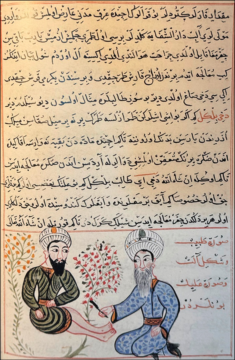
Figure 1 - Sabuncuoğlu treats the Dracunculus medinensis parasite surgically. (his own illustration in Cerrahiyetü’l Haniyye).
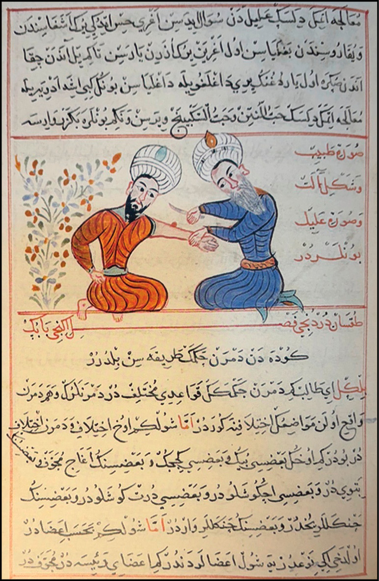
Figure 2 - Sabuncuoğlu treats painful subcutaneous parasites surgically. (his own illustration in Cerrahiyetu’l-Hanniyye).
IMPERIAL SURGERY (CERRAHIYETÜ’L HANIYYE)
There are three known copies of Cerrahiyetü’l Haniyye: one housed at the Fatih Millet Library in Istanbul, one at the Department of the History of Medicine at Istanbul University’s Çapa Faculty of Medicine, and one preserved in the Bibliothèque Nationale in Paris. The present analysis focuses specifically on the Paris manuscript of this work.
Imperial Surgery is divided into three sections, each generally organized as case reports presenting patients and their diseases. Some subsections provide detailed descriptions of the procedures performed, the surgical instruments employed, and the positioning of the patient, accompanied by vivid miniature illustrations that elucidate the procedures.
The first section of the book describes diseases and their treatments, with particular attention to cauterization techniques, the instruments used, and the sites of application. The second section presents surgical treatments of various diseases, including detailed explanations of surgical tools and types of incisions. The third section addresses the management of bone fractures and joint dislocations.
DISEASES CAUSED BY CUTANEOUS PARASITES
Diseases caused by cutaneous parasites are described in the second section in three successive parts, accompanied by two miniature illustrations.
In the first part, which addresses cutaneous parasites, the disease associated with Dracunculus medinensis is designated as the “Medina type.” The text clearly defines the clinical features of the disease, its causative organism, and its treatment, supplemented by a miniature depiction (Figure 1). Sabuncuoğlu states: “The treatment of the disease they call the Medina type is recorded. …The disease referred to as the Medina type is actually a vein, which may appear in the calves or in other limbs. It occurs in people living in very hot climates such as the Arabian lands, the Hijaz, and Abyssinia, where individuals are weak and lean. Just as worms may lodge in the intestines, small worms may also infest the space between the skin and the muscle, manifesting as redness and inflammation. In patients, the calf first becomes red, then the area heats intensely, followed by swelling, and eventually the tip of the ‘vein,’ resembling a root of grass, emerges from the swollen site. …When the tip of this vein appears, a small lead pellet is attached. The weight of the lead varies, depending on the thickness of the ‘vein,’ from one to two dirhams (1 dirham = 3.207 grams = 0.0070702247 pounds). Once tied, the lead hangs, and as the worm elongates, it winds itself around the lead. If it does not emerge completely, it re-enters the skin from its tip and causes the disease anew. In some patients, this worm measures one span in length, while in others it may reach up to ten spans. I recount this for the instruction of physicians. At one time, among the patients brought from Damascus, there were those infected with the Medina type... Three spans were extracted from one patient and twenty spans from another. If the worm breaks during extraction, a probe is to be inserted at the point of emergence and the site incised to remove whatever remains inside. This disease produces multiple swellings of the feet and toes in some individuals, and it creates many perforations in the foot in others.”
In the second part, another disease caused by filarial parasites is described, referred to as the “disease of vision.” The text explains: “In some regions, this illness is called the disease of sight. …In Turkish, it is caused by a worm known as İmic. This worm settles beneath the skin, wandering between the muscle and skin, moving upward and downward in the body, traveling from one limb to another until it eventually perforates the skin and emerges. This disease is caused by small worms. …These worms migrate, ascend toward the head, and may strike the eye, blinding it. … The worm must be removed to treat this illness. If extraction is not possible, both its anterior and posterior ends are tied, and the skin above it is incised to remove the worm. If the worm has penetrated into the muscle and cannot be located, then the area is cauterized. If the worm has ascended to the head and is advancing toward the eye, then the patient’s forehead is tightly bound, the area above the worm is cut, and the worm is excised.”
In the third part, Sabuncuoğlu discusses the treatment of a painful skin parasite (referred to as nafir) affecting the limbs, presented as a case study accompanied by a miniature illustration (Figure 2). The paper reads as follows: “...The ailment they call nafir is painful; it passes from one limb to another... I rolled up the patient’s sleeve, and along the line extending from the elbow to the middle finger, there was a slight swelling at the level of the wrist. After a while, as if mercury were flowing, the worm moved from the wrist toward the shoulder; the pain in the wrist shifted to the shoulder, and later, after some time, it passed to the side of the torso... If you wish to treat this disease, you should bind both above and below the site of the pain, excise the affected area, and then cauterize it...”
DiSCUSSiON
Before the 18th century, knowledge of cutaneous parasites in historical sources was largely based on interpretation. The Ebers Papyrus, dated to around 1500 BC, contains instructions for treating swellings of the limbs known as aat. These passages seem to allude to the nature of Dracunculus medinensis infection and to techniques for extracting the worm. This interpretation has been widely accepted by most parasitologists [8, 9]. However, since the term aat simply denotes “swelling,” the inference that this specific text describes cutaneous parasites remains speculative. Some parasitologists have argued that the “fiery serpents” afflicting the Israelites in the Red Sea region after the Exodus were in fact Dracunculus medinensis [10]. This interpretation was first advanced in the 1855 textbook by the parasitologist, theologian, and Hebraist Gottlob Friedrich Heinrich Küchenmeister, later translated into English under the title Animal and Vegetable Parasites [11]. In Arabic medical literature, references to the “Medina vein” have led some historians to suggest that Arab physicians may have regarded the worm as a decayed vein rather than a living parasite [9, 12].
Surprisingly, despite the well-documented clinical similarities in the subcutaneous migration of filarial worms and Dracunculus, as both can affect the eyes and cause parasitic infections resulting in visual impairment or blindness, no reliable early record of such cases was identified. A 1598 engraving was once thought to depict the extraction of a worm from the eye; however, this interpretation has been comprehensively debated, and the image is now generally considered to represent judicial punishment rather than a medical procedure [13]. The first unequivocal clinical report dates to 1770, when the French surgeon Mongin, working in Santo Domingo in the Caribbean, described a worm traversing a woman’s eye and recounted his unsuccessful attempt to remove it [14].
In the 15th-century treatise Imperial Surgery, Dracunculus medinensis disease was explicitly described, noting that the condition arises from organisms resembling larvae within the intestines and that its designation as “Irkı Medine,” or “the Medinan type,” is recorded. The text provides detailed information on the parasite’s length and the method of extraction. Furthermore, it discusses the type of surgical procedure to be performed in cases where extraction is not possible. The parasite responsible for blindness, along with the disease itself, appeared for the first time in history in Sabuncuoğlu’s study. As in many other areas of medicine, Imperial Surgery sheds light on the history of parasitic diseases through its descriptions of surgical interventions. Without interpretive ambiguity, the treatise presents skin parasites openly and, for the first time, correlates the causative organisms (larvae) with the clinical features of the diseases they produced.
Sabuncuoğlu is regarded as one of the most significant physicians in the history of medicine. His book, Imperial Surgery, was rediscovered in 1936 by the medical historian Süheyl Ünver [15]. With its rediscovery, the text secured its rightful place in the history of medicine for its contributions to neurosurgery, orthopedics, gynecology, pediatrics, and craniofacial surgery [16-20]. The miniatures contained in the manuscript are striking. Diseases are described in detail, accompanied by case presentations and clinical findings, while surgical procedures are visually illustrated. The miniatures faithfully reflect the text through scene construction, compositional arrangement, the identification of figures, and numerous other details. They avoid indirect narration or abstraction; instead, they aim to convey the subject matter to the reader in its entirety. The illustrations employ a distinctive two-dimensional technique, devoid of perspective, utilizing free colors independent of luministic art. In this way, surgical procedures are concretized. The deliberate absence of spatial depth in the miniatures focuses attention on the surgical act itself, conveying the intended message with unembellished simplicity.
Sabuncuoğlu presented a wide range of diseases and their treatments in a style that was both imaginative and unique, employing an academic register that transcended the limitations of his era. His command of Arabic, Persian, and Greek allowed him to clearly understand the foundational medical sciences of his time. The evidence in Imperial Surgery clearly demonstrates that, as early as the fifteenth century, physicians possessed knowledge and experience regarding parasitic diseases of the skin. Until recently, the historical record of such conditions had relied largely on interpretive, uncertain accounts. However, Cerrahiyetü’l-Haniyye, written by Sabuncuoğlu, shows that, in 15th-century Anatolia, the etiology, clinical presentation, and surgical treatment of cutaneous parasitic diseases were already understood in detail.
Conflict of interest
None
Funding
The author does not have a financial interest in any of the products, devices, or drugs mentioned in this article. No funding was received. All figures in this manuscript are reproductions of 16th-century historical illustrations that are in the public domain and do not require permission for reproduction.
REFERENCES
[1] Cox FE. History of human parasitology. Clin Microbiol Rev. 2002; 15 (4): 595-612.
[2] Bryan, C. P. The Papyrus Ebers (translated from the German). Geoffrey Bles, London, United Kingdom. 1930: 197.
[3] Jones, W. H., and E. T. Withington. Works of Hippocrates. Loeb Classical Library, Heinemann, London, United Kingdom. 1948-1953.
[4] Spencer, W. Translation of A Celsus De medicina. Loeb Classical. Library, Heinemann, London, United Kingdom. 1948-1953.
[5] Kuhn, K. (ed.). Galen (Galenus). Opera omnia, 22 vols. Leipzig, Germany. 1821-1833.
[6] Grove, D. I. A history of human helminthology. CAB International, Wallingford, United Kingdom. 1990.
[7] Linnaeus, C. Systema Naturae [System of Nature], 10th edn. Leiden. 1758.
[8] Foster, W. D. A history of parasitology. Livingstone, Edinburgh, United Kingdom. 1965.
[9] Grove, D. I. A history of human helminthology. CAB International, Wallingford, United Kingdom. 1990.
[10] The Holy Bible, New International Version (NIV). Zondervan. 1984; 21: 6-9.
[11] Ku¨chenmeister, F. Animal and vegetable parasites. Translated from the German by Edwin Lankaster. The Sydenham Society, London, United Kingdom. 1857.
[12] Löwy I. The Wellcome Trust illustrated history of tropical diseases. Med Hist. 1997; 41(4): 502-503.
[13] Gruntzig, J., and B. Jennes. Historical note on Loa loa: a reinterpretation. Am. J. Trop. Med. Hyg. 1977; 26: 697-683.
[14] Mongin. Sur un ver trouve´ sous la conjunctive a` Maribarou, isle Saint-Dominique. J. Med. Chir. Pharm. 1770; 32: 338-339.
[15] Unver S. Serefeddin Sabuncuoglu: Kitabul Cerrahiye-i Il- haniye. Istanbul, Turkey: Kenan Yayinlari. 1939.
[16] Keskil S, Sabuncuoglu H. Endoscopy in the 15th century. Minim Invasive Neurosurg. 2002; 45: 45-46.
[17] Sarban S, Aksoy S, Uzel I, et al. Orthopaedic techniques of Sabuncuog˘lu in the 15th century Ottoman period. Clin Orthop Relat Res. 2005; 439: 253-259.
[18] Kafali H, Aksoy S, Atmaca F, et al. Colored illustrations of obstetrics manipulations and instrumentation techniques of a Turkish surgeon Serafeddin Sabuncuoglu in the 15th century. Eur J Obstet Gynecol Reprod Biol 2002; 105: 197-202.
[19] San I, Oguz H, Kafali H. Colored illustrations of pediatric otorhinolaryngologic surgical techniques of a Turkish surgeon, Serefeddin Sabuncuoglu, in the 15th century. Int J Pediatr Otorhinolaryngol, 2005; 69: 885-891.
[20] Doğan F, Tutak FN. Craniomaxillofacial Surgery Techniques of 15th Century Turkish Surgeon Serefeddin Sabuncuoglu. J Craniofac Surg. 2021; 32(3): 828-831.