Le Infezioni in Medicina, n. 3, 366-370, 2026
CASE REPORTS
When enteritis reaches the heart: acute Campylobacter jejuni–associated perimyocarditis in a young adult
Margherita Scapaticci1, Monica Vignoli1, Sebastiano Nigro1, Mauro Colletta2, Rita Mancini1, Andrea Bartolini1
1LUM – Laboratory Medicine, AUSL Bologna, Bologna, Italy;
2Division of Cardiology, Maggiore Hospital, AUSL Bologna, Bologna, Italy
Article received 22 June 2026 and accepted 19 August 2026
Corresponding author
Andrea Bartolini
E-mail: andrea.bartolini@ausl.bologna.it
Summary
Campylobacter jejuni is a common cause of bacterial gastroenteritis, while cardiac involvement is a rare extraintestinal complication. We report the case of a 21-year-old immunocompetent man who developed acute perimyocarditis three days after microbiologically confirmed C. jejuni enteritis. He presented with pleuritic chest pain, elevated inflammatory markers, and increased high-sensitivity troponin I levels. Echocardiography showed preserved ventricular function, whereas cardiac magnetic resonance imaging (CMR) demonstrated myocardial edema and subepicardial-to-mid-myocardial late gadolinium enhancement, fulfilling the Revised Lake Louise Criteria for acute myocarditis. The patient had an uncomplicated clinical course and was discharged in stable condition. This case highlights the importance of considering perimyocarditis in young patients presenting with chest pain following recent gastroenteritis and underscores the diagnostic value of CMR.
Keywords: Campylobacter jejuni; perimyocarditis; cardiac magnetic resonance imaging; extraintestinal complications; gastroenteritis.
INTRODUCTION
Campylobacter jejuni is one of the most common bacterial causes of acute gastroenteritis worldwide, typically acquired through the consumption of contaminated food or water [1, 2]. Although the infection is usually self-limiting, a variety of extraintestinal manifestations have been described, including reactive arthritis, Guillain–Barré syndrome, bacteremia, and, less frequently, cardiac involvement [3-8].
Acute myocarditis and perimyocarditis associated with C. jejuni infection are uncommon but potentially serious complications. Most reported cases have occurred in young, otherwise healthy individuals and typically develop within days of the onset of gastrointestinal symptoms [9-14]. Because the clinical presentation may mimic other causes of acute chest pain, early recognition is essential to ensure appropriate diagnostic evaluation and management. We report the case of a 21-year-old immunocompetent male who developed acute perimyocarditis following a recent C. jejuni infection.
PATIENT HISTORY
The study was conducted in accordance with the principles of the Declaration of Helsinki. Written informed consent for publication of clinical data was obtained from the patient.
The patient initially presented to the emergency department (ED) with a five-day history of abdominal pain and diarrhea, associated with fever, dehydration and mild hypokalemia. Laboratory investigations showed an elevated C-reactive protein (CRP) level of 21.79 mg/dL (reference limit <0.5 mg/dL), and stool samples were collected for microbiological investigation. Intravenous rehydration and empirical ceftriaxone were administered, and the patient was discharged after clinical improvement on empirical oral cefixime (400 mg once daily for five days).
Three days later, the patient returned to the ED complaining of acute pleuritic chest pain radiating to the left arm, which worsened with positional changes. Diarrhea had resolved, although stools remained poorly formed, and he was afebrile and hemodynamically stable.
Past medical history was unremarkable, with no previous cardiovascular disease or surgical interventions. No drug allergies were reported. Family history was notable for premature coronary artery disease, as the patient’s father had suffered a myocardial infarction at the age of 52 years.
Electrocardiogram (ECG) showed sinus rhythm at 71 bpm and was otherwise within normal limits. Chest X-ray was unremarkable.
Laboratory tests demonstrated significant inflammation and myocardial injury, with a CRP level of 3.28 mg/dL and markedly elevated high-sensitivity troponin I levels (>1000 ng/L on point-of-care testing; 99th percentile upper reference limit: 25.7 ng/L) subsequently confirmed at 7402.4 ng/L by central laboratory analysis (99th percentile upper reference limit: 19.8 ng/L for males). Renal function, complete blood count and serum electrolytes were within reference ranges.
Review of the microbiological investigations requested during the patient’s previous ED visit for acute gastroenteritis revealed growth of Campylobacter jejuni in stool culture after 48 hours of incubation. Stool cultures were otherwise negative for Salmonella spp. and Shigella spp., while stool antigen assays for enteric adenovirus and C. jejuni and serological tests for Brucella spp. and Salmonella typhi were negative. In view of the improvement of the gastrointestinal illness, no targeted antimicrobial therapy against C. jejuni was initiated after microbiological identification. The patient was subsequently admitted to the coronary care unit (CCU) for further evaluation and monitoring because of the clinical presentation and laboratory findings.
Upon admission to the CCU, the patient was alert, oriented, and cooperative. He was without ongoing chest pain and dyspnea at rest and was breathing comfortably on room air. Physical examination revealed normal vesicular breath sounds bilaterally, without crackles or other adventitious sounds. No peripheral edema was observed.
The ECG showed sinus rhythm at 71 bpm, with a normal PR interval and nonspecific repolarization abnormalities.
Transthoracic echocardiography performed on the same day demonstrated a non-dilated left ventricle (end-diastolic diameter 47 mm; end-diastolic volume 102 mL) with preserved global systolic function (LVEF 55%) and circumscribed hypokinesia of the mid-inferior wall. Global longitudinal strain was normal (-21%). Mild mitral regurgitation was present. The left atrium was of normal size (indexed left atrial volume 23 mL/m²). The aortic root was not dilated (25 mm). Diastolic function was normal, with an E/e′ ratio of 5. Right heart chambers were within normal limits, with preserved right ventricular systolic function (TAPSE 26 mm). Mild tricuspid regurgitation was noted, with an estimated systolic pulmonary artery pressure of 25 mmHg. No pericardial effusion was detected.
During CCU admission, continuous cardiac monitoring revealed no arrhythmias or hemodynamic instability. Ramipril (1.25 mg twice daily) was initially administered during the early phase of cardiological management. Serial troponin measurements showed a progressive decline, and after three days, following clinical stabilization, the patient was transferred to the cardiology ward, where cardiac magnetic resonance (CMR) imaging was performed. Following completion of the diagnostic work-up and confirmation of preserved left ventricular systolic function, ramipril was progressively reduced and discontinued before discharge.
CMR revealed normal left ventricular size and preserved biventricular systolic function (LVEF 56%; RVEF 54%), with mild hypokinesia of the basal inferolateral wall. T2-weighted sequences demonstrated myocardial edema in the basal inferolateral wall, with increased native T1 and T2 values and corresponding subepicardial-to-mid-myocardial late gadolinium enhancement. These findings fulfilled the Revised Lake Louise Criteria [15]. A small circumferential pericardial effusion, not detected on the initial echocardiography, was also present, without pathological pericardial thickening or contrast enhancement. Representative CMR images are shown in Figure 1.
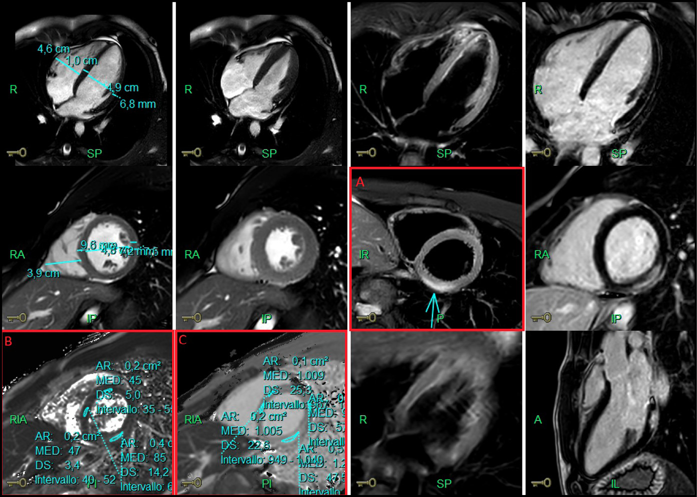
Figure 1 - Representative cardiac magnetic resonance images obtained during the acute phase. The red-highlighted panels show the main tissue-characterization findings: (A) short-axis late gadolinium enhancement image demonstrating focal subepicardial-to-mid-myocardial enhancement of the basal inferolateral wall (arrow); (B) T2 mapping showing an increased T2 relaxation time in the same region (85 ms; reference value 48 ± 3 ms), consistent with myocardial edema; and (C) native T1 mapping showing an increased T1 relaxation time in the basal inferolateral wall (1200 ms; reference value 983 ± 26 ms).
The remainder of the hospital course was uneventful, with no recurrence of chest pain, no signs or symptoms of heart failure, and no arrhythmias detected on continuous telemetry monitoring.
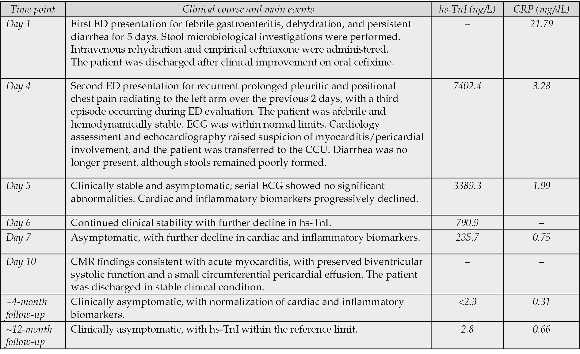
At discharge, the patient was free of chest pain and dyspnea at rest, afebrile, eupneic on room air, and hemodynamically stable, with a blood pressure of 110/60 mmHg. Physical examination revealed normal vesicular breath sounds bilaterally, without crackles. Cardiac auscultation showed a regular rhythm with normal heart sounds and a grade 2/6 systolic murmur. No peripheral edema was present. He was advised to refrain from strenuous physical activity for 6 months and was scheduled for outpatient cardiology follow-up. At approximately 4 months, the patient remained clinically asymptomatic, with normalization of hs-TnI (<2.3 ng/L) and CRP (0.31 mg/dL). At approximately 12 months, he remained asymptomatic and hs-TnI was still within the reference limit (2.8 ng/L). Table 1 summarizes the clinical course and serial laboratory findings.
Table 1 - Clinical course and serial laboratory findings. Timeline of the main clinical events and serial high-sensitivity troponin I (hs-TnI) and C-reactive protein (CRP) values during the acute phase and follow-up. Laboratory values shown were obtained by central laboratory analysis. The 99th percentile upper reference limit for hs-TnI was 19.8 ng/L in males, and the CRP reference limit was <0.5 mg/dL. CCU, coronary care unit; CMR, cardiac magnetic resonance; CRP, C-reactive protein; ED, Emergency Department; hs-TnI, high-sensitivity troponin I.
DISCUSSION
Cardiac involvement is an uncommon extraintestinal manifestation of C. jejuni infection. Although Campylobacter spp. are among the leading causes of bacterial gastroenteritis worldwide, myocarditis and perimyocarditis have been only occasionally reported. Available evidence suggests that affected patients are predominantly young, otherwise healthy males who develop cardiac symptoms within a few days of the onset of gastrointestinal illness. Clinical presentation is typically characterized by acute chest pain, elevated inflammatory markers, and increased cardiac troponin levels [9-14].
The pathophysiological mechanisms underlying Campylobacter-associated perimyocarditis remain incompletely understood [16]. Because bacteremia occurs in fewer than 1% of Campylobacter infections, direct myocardial invasion is considered unlikely in most cases. Current evidence favors an immune-mediated process, potentially driven by molecular mimicry and systemic inflammatory activation, similar to the mechanisms implicated in other post-infectious complications of Campylobacter infection [17]. The close temporal relationship between enteritis and cardiac involvement observed in our patient is consistent with this hypothesis.
In young patients presenting with chest pain and elevated cardiac biomarkers, the differential diagnosis includes acute coronary syndrome, viral myocarditis, Takotsubo cardiomyopathy, and isolated acute pericarditis [3, 15, 18-19]. In our case, an ischemic etiology was considered unlikely because electrocardiography showed no ischemic changes, and CMR demonstrated no resting myocardial perfusion defects and a non-ischemic pattern of myocardial injury, characterized by subepicardial-to-mid-myocardial involvement of the basal inferolateral wall [15]. Echocardiography showed preserved global left ventricular systolic function despite limited regional hypokinesia. Takotsubo cardiomyopathy was considered unlikely because the pattern of regional wall motion abnormalities was not typical and CMR demonstrated focal inflammatory myocardial tissue abnormalities. Although viral myocarditis could not be completely excluded, the close temporal association with microbiologically confirmed C. jejuni enteritis, together with the absence of a more likely alternative etiology, was considered suggestive of Campylobacter-associated perimyocarditis.
CMR played a pivotal role in the diagnostic work-up. As the reference non-invasive imaging modality for suspected myocarditis, it is recommended by the latest ESC Guidelines on Myocarditis and Pericarditis for myocardial tissue characterization and enables the detection of myocardial edema and non-ischemic patterns of myocardial injury [18]. In our patient, the presence of myocardial edema and subepicardial-to-mid-myocardial late gadolinium enhancement fulfilled the Revised Lake Louise Criteria for acute myocarditis, supporting the diagnosis [15]. Similar imaging findings have been reported in previous cases of Campylobacter-associated myocarditis and perimyocarditis, comprehensively reviewed by Hessulf et al. and more recently updated by Zouganeli et al. [9-10]. These reports describe a remarkably consistent clinical presentation, with young, otherwise healthy patients developing acute chest pain within a few days of Campylobacter enteritis, associated with elevated cardiac biomarkers and non-ischemic CMR findings. Our findings are in keeping with this previously described clinical pattern.
CONCLUSIONS
C. jejuni–associated perimyocarditis is an uncommon but clinically relevant complication of bacterial gastroenteritis. Cardiac involvement should be considered in patients presenting with chest pain shortly after enteric infection, particularly when Campylobacter infection has been microbiologically confirmed. Early recognition and timely CMR evaluation are essential for establishing the diagnosis, excluding alternative etiologies, and guiding appropriate management.
Conflicts of interest
The authors declare no conflict of interest.
Funding
Nothing to declare.
REFERENCES
[1] European Food Safety Authority. https://www.efsa.europa.eu/en/topics/topic/campylobacter (last access: 16 June 2026).
[2] Shane AL, Mody RK, Crump JA, et al. Infectious Diseases Society of America Clinical Practice Guidelines for the Diagnosis and Management of Infectious Diarrhea. Clin Infect Dis. 2017; 65(12): 1963-1973.
[3] Uzoigwe C. Campylobacter infections of the pericardium and myocardium. Clin Microbiol Infect. 2005; 11(4): 253-255.
[4] Pacanowski J, Lalande V, Lacombe K, et al. Campylobacter bacteremia: clinical features and factors associated with fatal outcome. Clin Infect Dis. 2008; 47(6): 790-796.
[5] Pope JE, Krizova A, Garg AX, Thiessen-Philbrook H, Ouimet JM. Campylobacter reactive arthritis: a systematic review. Semin Arthritis Rheum. 2007; 37(1): 48-55.
[6] McCarthy N, Giesecke J. Incidence of Guillain-Barré syndrome following infection with Campylobacter jejuni. Am J Epidemiol. 2001; 153(6): 610-614.
[7] Scallan Walter EJ, Crim SM, Bruce BB, Griffin PM. Incidence of Campylobacter-Associated Guillain-Barré Syndrome Estimated from Health Insurance Data. Foodborne Pathog Dis. 2020; 17(1): 23-28.
[8] Fischer GH, Hashmi MF, Paterek E. Campylobacter Infection. Treasure Island (FL): StatPearls Publishing. 2024.
[9] Hessulf F, Ljungberg J, Johansson P-A, Lindgren M, Engdahl J. Campylobacter jejuni-associated perimyocarditis: two case reports and review of the literature. BMC Infect Dis. 2016; 16: 289.
[10] Zouganeli V, Kourek C, Bistola V, et al. Campylobacter jejuni-Related Myocarditis: A Case Report and Review of the Literature. J Clin Med. 2024; 13(24): 7551.
[11] Moffatt CR, Moloi SB, Kennedy KJ. First case report of myopericarditis linked to Campylobacter coli enterocolitis. BMC Infect Dis. 2017; 17(1): 8.
[12] Gomes SA, Trigo C, Pinto FF. Campylobacter coli myocarditis: a case report. Cardiol Young. 2021; 32(7): 1172-1174.
[13] Elford A, Thakkar H, MacIntyre P. The way to a man’s heart is through his stomach: a curious case of myopericarditis associated with Campylobacter enteritis. Aust J Gen Pract. 2021; 50(5): 305-307.
[14] Chantzaras AP, Karageorgos S, Panagiotou P, et al. Myocarditis in a Pediatric Patient with Campylobacter Enteritis: A Case Report and Literature Review. Trop. Med. Infect. Dis. 2021; 6(4): 212.
[15] Ferreira VM, Schulz-Menger J, Holmvang G, et al. Cardiovascular Magnetic Resonance in Nonischemic Myocardial Inflammation: Expert Recommendations. J Am Coll Cardiol. 2018; 72(24): 3158-3176.
[16] Young KT, Davis LM, Dirita VJ. Campylobacter jejuni: molecular biology and pathogenesis. Nat Rev Microbiol. 2007; 5(9): 665-679.
[17] Suliman BA. Potential clinical implications of molecular mimicry-induced autoimmunity. Immun. Inflamm. Dis. 2024; 12: e1178.
[18] Schulz-Menger J, Collini V, Gröschel J, et al. ESC Scientific Document Group. 2025 ESC Guidelines for the management of myocarditis and pericarditis. Eur Heart J. 2025; 46(40): 3952-4041.
[19] Lyon AR, Bossone E, Schneider B, et al. Current state of knowledge on Takotsubo syndrome: a Position Statement from the Taskforce on Takotsubo Syndrome of the Heart Failure Association of the European Society of Cardiology. Eur J Heart Fail. 2016; 18(1): 8-27.